The Site for Healthcare Professionals: 2021
WE DO ADVERTISEMENT SERVICES FOR YOUR HEALTHCARE PRODUCTS, PROMOTING YOUR HEALTHCARE EVENTS, TECHNICAL REVIEWS FOR YOUR MEDICAL DEVICES, ETC...
PLEASE CONTACT US FOR FURTHER DETAILS ABOUT OUR SUPPORTS.....
OUR EMAIL ADDRESS:- sam.gastondiaz@gmail.com

Thursday, September 30, 2021

ABG Machine

 Introduction:
The amount of arterial gases, such as oxygen and carbon dioxide, is measured in an arterial blood gas (ABG) test.

In whole blood specimens, analyzers are utilized to assess blood gas, pH, electrolytes, and certain metabolites. They can measure pH, carbon dioxide, and oxygen partial pressures, as well as the concentrations of a variety of ions (such as sodium, potassium, chloride, bicarbonate) and metabolites (e.g.: calcium, magnesium, glucose, lactate).

ABG analysis is also used to detect acidosis and alkalosis conditions, kidney and lung function, response to treatment, and acid-base balance in the body.


Overview:
Some of the attributes of ABG machines covered in this article include: 
  • what an ABG machine is
  • what it is used for
  • where it is utilized
  • what the risks are
  • advantages and disadvantages of ABG machine systems.


What is an ABG machine?
An ABG (Arterial-Blood Gas) machine, also known as Arterial-Blood Gas Analyzer, is used to measure the amount of oxygen and carbon dioxide (called blood gases) in the blood. It is also used to determine the blood pH (Power of Hydrogen). 


What is a Blood gas Test?
The amount of oxygen and carbon dioxide in the blood is measured by a blood gas test. It can also be used to determine the pH, or acidity, of the blood.  A blood gas analysis or arterial blood gas (ABG) test is the most popular name for the test. Throughout the body, red blood cells deliver oxygen and carbon dioxide. They are known as 'Blood gases'. When blood travels through the lungs, oxygen enters the bloodstream, while carbon dioxide exits the bloodstream and enters the lungs. The blood gas test can show how successfully the lungs exchange oxygen into the bloodstream and eliminate carbon dioxide from the blood.


Why is the ABG machine used?
  • The ABG machine is used to diagnose several breathing and lung conditions such as chronic obstructive pulmonary disease (COPD), or cystic fibrosis (a life-threatening hereditary disorder that affects the lungs and digestive system).
  • To check if the blood requires extra oxygen or any assistance with breathing.
  • To check the acid-base balance present in the blood. There can be too much acid in case of kidney failure, infection, or any toxic component intake.
  • To monitor whether and how the lungs are responding to treatment.
  • To monitor if the lungs are removing the right amount of carbon dioxide (CO2).
  • To monitor vital kidney functions.
  • To find the presence of respiratory acidosis, respiratory alkalosis, metabolic acidosis, or metabolic alkalosis.


How does the ABG machine work? 
  • At 37°C, blood gas analyzers have three electrodes that measure pH, PCO2, and PO2. They were first introduced in the 1960s, after innovations by R Stow (CO2) and L Clark (PO2) in 1954.
  • Electrodes are used in blood gas/pH analyzers to determine pH, carbon dioxide partial pressure, and oxygen partial pressure in the blood. 
  • A dry reagent pad system is used in chemistry analyzers, in which a filter pad impregnated with all of the chemicals required for a certain reaction is placed on a thin plastic strip. 
  • The ion-selective electrode (ISE) approach is used in electrolyte analyzers, in which measurements of ion activity in a solution are made potentiometrically using an external reference electrode and an ISE with an internal reference electrode.


How is blood prepared for an ABG test?


The only anticoagulant used to prepare blood samples for blood-gas analysis is Heparin. Inadequate anticoagulation and the production of tiny blood clots, which can block the sample stream of blood gas analyzers and invalidate results, are two of the most common practical difficulties associated with blood gas analysis.


How is an ABG machine operated?


Whole blood samples are put into the analyzer in tubes, reaction cuvettes (a piece of laboratory equipment designed to contain samples for spectroscopic analysis), syringes, capillaries, or test strips. It is critical to remember that no blood should be injected or poured into the machine. Using a keypad or a linked computer, the operator can pick the tests that will be performed on the sample.


Where is the ABG machine used?
Hospital, patient bedside, physician's office, clinical laboratory, and home are some of the places where this device is used.


How is the blood drawn for the ABG test? 
Blood is usually drawn from an artery. Blood from a vein may be used in some instances (venous blood gas).


Blood is most typically drawn from one of the following arteries:
• The radial artery (wrist)
• The brachial artery (arm)
• The femoral artery (groin) 

Before collecting a blood sample from the wrist area, the health care professional may do a circulation test, also known as Allen Test (a test that determines whether or not the blood flow to the hand is normal. For a few seconds, the health professional drawing blood will apply pressure on the arteries present in the wrist. The blood flow to the hand will be cut off temporarily, and the hand will turn cool and pale).

A tiny needle is inserted through the skin into the artery by the provider. The sample is sent to a laboratory for analysis right away.


What are the main components of an ABG?
There are six main components to an ABG. They are as follows:
1. pH
2. Oxygen partial pressure (PaO2)
3. Carbon dioxide partial pressure (PaCO2)
4. Bicarbonate (HCO3)
5. Oxygen saturation (O2 Sat)
6. Oxygen concentration (O2 CT)


What are some of the body conditions that require ABG analysis?
The ABG machine is used to diagnose several breathing and lung conditions. Some of the common conditions ABG is used for are:

1. Respiratory acidosis:
Inadequate alveolar ventilation causes respiratory acidemia (acidosis), which results in the retention of carbon dioxide and an increase in free hydrogen ions. It happens when reduced breathing (hypoventilation) raises the concentration of carbon dioxide in the blood and lowers the pH of the blood.

2. Respiratory alkalosis:
Respiratory alkalosis is caused mainly by excessive carbon dioxide excretion/exhalation (hyperventilation), which results in the exhalation of more CO2 than is normal. As a result, the number of free hydrogen ions decreases, resulting in an alkaline condition.

3. Metabolic acidosis:
Excess fixed acid production, such as lactate or HCO3 loss, is a symptom of metabolic acidosis. It occurs when the body produces too much acid or when the kidneys do not remove enough acid from the body. Acidemia (acidosis) is characterized by a low blood pH (7.35) caused by the body's excessive production of hydrogen ions or its inability to produce bicarbonate (HCO3) in the kidneys.

4. Metabolic alkalosis:
Metabolic alkalosis is caused by a decrease in hydrogen ion concentration, which leads to an increase in bicarbonate, or is caused by increased bicarbonate concentrations.


How are the ABG results interpreted?
The pH should be checked first to see if the patient is within normal range, above the normal range, or below the normal range. A patient is in alkalosis if his or her pH is more than 7.45. If the pH is below 7.35, the patient is in acidemia (acidosis). Remember that the lower the pH number, the more acidic the body is. Even though the pH is normal, acidosis or alkalosis may still exist since the body can compensate to keep the pH balanced.

After that, examine the PaCO2 level. This will reveal if the variations in blood gas are caused by the respiratory system or by metabolic alterations. The medical expert will be able to fully interpret the blood gas when combined with the HCO3.

Below is a table to help in better understanding of ABG results interpretation:

ABG                                    

             pH

PaCO2

HCO3

Respiratory Acidosis


             Low

        
 High

Normal

Respiratory Alkalosis


             High

 Low

Normal

Metabolic Acidosis


             Low

Normal


 Low

Metabolic Alkalosis


             High

Normal


 High


· What are the normal ABG values?

pH

7.35-7.45

pCO2

35 mmHg – 45 mmHg

pO2

75 mmHg – 100 mmHg

HCO3-

22 mEq/L – 26 mEq/L

O2 Saturation

> 95 %

                             *mEq/L – milliequivalents per litre             *mmHg – millimetre of mercury


The normal value varies depending on the laboratory, machine model, and age factor (for example, infants may show different normal values compared to the elderly).


What are the risks associated with ABG?
A blood sample collected from an artery has a low risk of causing complications. Although sometimes, any test that requires the use of a needle entails the risk of bleeding, infection, and bruise. There may be some tenderness/soreness where the needle was inserted.
  • There could be a tiny bruise on the affected area. It is advised to apply pressure on the location for at least 10 minutes after the needle is removed to reduce the risk of bruising.
  • A nerve or an artery may be damaged by the needle in rare situations. The artery may become blocked as a result of this. 
  • When the process is performed appropriately, there is very little risk. The size of veins and arteries differs from person to person and from one side of the body to the other. It may differ because drawing blood from some patients can be more difficult than from others.

Other concerns of having blood drawn are minor, although they can include:
  • Feeling faint or lightheaded
  • Several punctures are used to locate blood vessels.
  • Hematoma - a type of hemorrhage (blood build-up under the skin)
  • Excessive loss of blood
  • Infection

What is the cost of an ABG machine?
In India, the price of an ABG Machine typically ranges from INR 3 lakh to INR 7 lakh.


What are the advantages of an ABG machine?
These ABG systems provide a number of advantages, including 
  • Less maintenance, 
  • Improved fault detection, and 
  • Reliable quality control. 
Another benefit is a reduction in phlebotomy-related blood loss, which is critical in settings such as the operating room (OR) or intensive care unit (ICU), where blood conservation is critical. Analyzers are becoming faster, smaller, and more user-friendly, and they are demonstrating the potential to perform accurate testing with smaller blood samples.


What are the disadvantages of an ABG machine?
  • One of the disadvantages includes a sample being mishandled or testing performed wrong. 
  • In a hospital, emergency department, or big clinical laboratory, qualified healthcare providers perform blood gas analysis. 
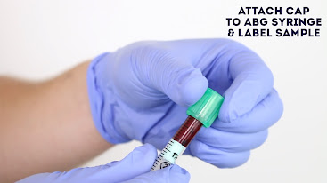
These tests are "stat" tests, which means they should be completed as soon as possible after the sample has been collected. The obtained blood sample for arterial blood gases (ABGs) degrades rather quickly. Therefore, if a testing delay is anticipated, the sample should be maintained on ice and rewarmed later for accurate analysis. If any air bubbles remain in the top of the syringe after the sample has been collected, they must be removed. The syringe is then placed on ice and transferred for quick analysis when the needle is sealed.

Some instances of the sample being mishandled are:
If the sample contains air bubbles.
If the sample gets contaminated with venous blood.
If the blood sample coagulates due to insufficient heparin coating of the tube.
If there is an excess of heparin (liquid heparin will dilute the blood and cause changes in the pH value, leading to wrong value display).
Occurrence of hemolysis before the sample being placed in the ABG analyzer.
Another disadvantage could be a lack of proper documentation, leading to the loss of important patient details.


What are the instrumental errors that can occur in an ABG machine?
  • The inaccurate temperature of electrolytes.
  • Improper calibration of ABG analyzer
  • Inadequate quality control and electrode maintenance (electrodes have various measurable lifespans).
  • Poorly compensated non-linearity of the Clark electrode.


*(CLARK ELECTRODE - a well-known sensor for measuring dissolved oxygen concentration in a water solution. This type of electrochemical sensor has the advantage of being easy to detect extremely low oxygen levels. This electrode needs to be replaced once every 3 years as its Teflon membrane becomes coated with proteinaceous filth).


How to troubleshoot instrumental errors?


Ensuring proper maintenance of the machine:-
Frequent and adequate calibration of ABG analyzer. Calibration is a procedure performed on blood gas electrodes prior to testing blood samples to ensure that readings are accurate within the expected range.

Gases used for calibration should be exceedingly precise and able to be traced back to certification from the National Institute of Standards and Technology (NIST):-
Initial adequate Quality control checking of the machine and its parts.

When it comes to blood gas electrodes, quality control is monitoring the instrument's performance on a regular basis to ensure calibration, stability, and reliability.

The accuracy and precision of blood gas measurements are assessed using statistical approaches. 

The most controllable part of quality assurance is certainly quality control.
The lifespan of the machine should be considered and appropriate measures should be taken. 
Internal quality control processes are meant to ensure that a laboratory's instruments 
      (e.g., in this case, electrodes) function as expected. 

They consist of standard methods and protocols for detecting irregularities in performance. 

Most external regulatory or accreditation agencies (e.g., Joint Commission for the Accreditation of Healthcare Organizations [JCAHO], Clinical Laboratories Improvement Amendments [CLIA]) demand internal quality monitoring.
• No particular instructions for routine calibration can be supplied due to the many varying processes, designs, and recommendations of various manufacturers. Therefore, operators must follow the manufacturer's guidelines to the letter.


Article Contributed By:-


Wednesday, September 29, 2021

C-Arm Machine.

 Introduction
A C-arm machine is a medical imaging device that works based on x-ray technology.  The name c arm machine is derived from a C-shaped arm that is used to connect the X-ray source and x-ray detector to one another. The first C-arm machine was introduced in 1955. As medical imaging has an important role in different diagnoses and treatment of patients. It has become important for medical practitioners in order to have a thorough understanding of many different complicated techniques exist. 

Since the introduction of the c arm machine, there are so many technologies are advanced. These machines are highly flexible and which is widely used in different medical practices. For instance, they are widely used during orthopedics, anesthesia, and various complicated surgeries. Nowadays, movable medical imaging systems are an important part of everyday life. 

Different specialists in the fields like surgery, orthopedics, vascular surgery, traumatology, and cardiology use c-arm machine for surgical operation imaging.  The c-arm machines provide high-resolution x-ray images in real-time medical imaging so that it allows the physician to monitor progress happen at any time during the operation and immediately make any corrections that might be necessary. As a result, the treatment will be good and the patient may recover very quickly.

What is a C-arm Machine?
C-arm machines are medical imaging devices which are work based on x-ray technology.  They are highly flexible and used in various OTs in hospitals. The name c- arm is derived from the c-shaped arm present in the device, which is used to connect the x-ray source and detector together. 


COMPONENTS OF C-ARM MACHINE
In general c-arm machine is made up of mainly three parts.

1. X-ray Generator:-
An x-ray generator is the main part of the c-arm machine which produces x-rays whenever an electrical current is applied to it.  Through an x-ray generator, the current is allowed to flow. An x-ray generator is a device that acts as the primary control mechanism for the entire c-arm machine. An x-ray generator is placed inside the frame where the c-arm is mounted and it can be directly controlled by the work station unit so that the operator can the operation of the system. 


2. Imaging system
One of the main parts of the c-arm machine is the imaging system. The imaging system of the c-arm machine has the ability to move in multiple directions in a single procedure. Due to this c-arm machines are highly advantageous during different surgical activities. Such as ortho, urology, and cardiology. The whole system is highly compact and has lightweight so that allows multiple movements in various directions.

3. Work Station
The workstation is the major part of the c-arm machine. The entire activity of the c-arm machine is controlled with the help of work station. So, the workstation is called the brain of c-arm machines.


WORKING PRINCIPLES OF C-ARM MACHINE
The C-arm machine consists of an x-ray generator and an x-ray image intensifier. An x-ray generator emits x-rays that penetrate the patient’s body. Then the image intensifier part changes the x-rays into a visible image which is displayed on the c-arm monitor. 


The c-shaped connecting element of the c-arm machine allows the movement of a machine horizontally, vertically, and around the swivel axes, so that x-ray images of the patient can be taken from any angle.

Applications of c-arm Machine  
  • During different surgeries, the real-time image visualization of internal organs like the liver, kidney, bone, and many different anatomical structures can be obtained.
  • C-arm systems of the c-arm machines can guide a needle placement procedure usually it is applicable during injecting Anastasia. Because they allow us to identify the exact place where the medication can be inserted.
  • C-arm machines are widely used during orthopedic, different complicated surgeries, and indifferent emergency procedures.

Types of c-arm Machine
1. Fixed c-arm/ Cath Lab:-
Fixed c arms also called Cath labs which produce highly detailed imaging with high generator output in KW.


2. Mobile c-arm
Mobile c-arm unit offers highly cost-effective imaging so that we can get enough images for every clinical procedure.


3.  Mini c-arm
They are like conventional c-arm used x-rays to provide images. But c-arm offers a better advantage than conventional c arm machines. It is a surgeon operated and it is not required to have a radiographer.
  • We will have better workplace access due to its small size. Because it is so simple to use and compact, it is popular for daily use, particularly for hand, ankle, and foot surgeries.
  • Better accuracy and enhanced maneuverability
  • Reduced delays and lower cost of surgery


Advantages of c-arm Machine
  • The major advantage of c-arm machines is their ability to move in different directions so that we can achieve the optimum angle for high-quality images.
  • C arm machines are widely used during complicated surgeries. If there is a need to place during surgery, c-arms are highly advantageous.

Disadvantages
The device can rotate in multi directions which leads it to offer poor image quality. 
When the device is put for a longer operation time it will prone to heating.
If the exposure of the eye to the machine increase it will cause some eye damages.
When we use vertical AP with a tube on top the high exposure will cause head and neck pain.
Trimming down the size of the c-arm machine leads to positioning and angulation limitations in the operating room.
                         

Use of the c-arm machine in orthopedic surgery
They are mainly used in studies that require maximum positional flexibility. This includes: -
  • General surgery, orthopedic procedures, and urology procedures
  • Vascular and neurology procedures
  • Cardiac procedures

Possible troubles arise in C-arm machine
  • During calibration, a grainy image will get displayed.
  • Image quality is also often affected by dust or lint particles in the Ⅱ or CCD camera. 
  • Charger fail error
  • X-ray tube may be pitted or the image intensifier might be old
  • Low mA error
  • Hard drives may fill or need replacing
  • Communication error
  • foreign objects may obscure the C-arm image.

Troubleshooting of C arm machine
  • In case of an unresponsive or faulty error in the image processing of the c arm machine which is a digital artifact, we have to unplug and reinsert the cables and boards.
  • For physical artifacts, we have to use a cleaning lens manually with an eyeglass cloth. If the problem persists, there is a good chance the artifact is lodged inside the camera.
  • If wiping the lens fails removing the image intensifier grid panel and cleaning behind the grid with an air puffer is enough solution for the artifact issues. If this also not working, we have to replace our machine.
  • Grainy or a high contrast image will occur when the c arm is positioned near different tissue densities such as the thoracic spine and lumbar spine which are caused when AEC (automatic exposure controls) tries to adjust to both densities. So, in this case, we have to center the tube to the densest area first and wait for AEC to adjust then move to the body part being imaged.


Article Contributed By:-


Monday, September 20, 2021

Electro - Convulsive Therapy (ECT)

INTRODUCTION:-


Electroconvulsive Therapy (ECT) is a treatment method used for people who suffer from severe depression or bipolar disorders. In this therapy, a small electric current is passed through the patient’s brain, which is under general anesthesia, for inducing a brief seizure in the brain. Inducing such seizures causes a change in brain chemistry that reduces the symptoms of some mental illness, slowly bringing back the patient to his/her original condition. ECT is preferred only when all other treatments seem to be unsuccessful.

In earlier days, people had negative feedback about ECT as it used high electric current without anesthesia, leading to many serious side effects such as memory loss and bone fractures. But now ECT is considered much safer, with less side effects as it uses a small electric current, that too with general anesthesia. ECT therapy is generally carried out by a group of trained medical professionals including a psychiatrist, anesthesiologist, and physician assistants.

HISTORY OF ECT THERAPY
ECT therapy is one of the oldest treatment methodology used in the field of psychiatry. Roman psychiatrists, Ugo Cerletti and Lucio Bini developed the first ECT machine in 1938. Patient diagnosed with Schizophrenia was first treated with this machine and it was found successful. Ugo Cerletti and Lucio Bini were awarded Nobel Prize in medicine in 1930s.

The idea of inducing epileptic seizures for treating patients was proposed by Meduna, a Hungarian neuropathologist and psychiatrist. In 1940, ECT was used worldwide and was considered an effective technique for treating severe mental illness. In the 1960s, psychiatrist Thomas Szasz pioneered the ‘anti-psychiatry movement’ which criticized psychiatry and considered ECT as inhumane and torturous. Due to this reason, ECT took a back seat during 1960-1970 but returned to the mainstream mode of treatment in the 1980s.


PSYCHIATRIC DISORDERS TREATED WITH ECT
ECT can provide significant improvement in certain mental illness conditions. ECT is used to treat:-
  • Major depression, including a desire to commit suicide.
  • Severe mania, it is a bipolar disorder that includes intense euphoria, agitation, or hyperactivity. Impaired decision-making, impulsive or risky behavior, substance abuse, and psychosis are other signs of mania.
  • Schizophrenia, it is a disorder that affects a person’s ability to think, feel and behave properly.
  • Catatonia, it is associated with Schizophrenia and some other psychiatric disorders and is characterized by lack of movements, lack of speech, fast or strange movements, etc...
  • Treatment-resistant depression, depression that doesn’t get improved with other treatments.

TYPES OF ECT THERAPY

There are two types of ECT:
  • Bilateral ECT: In bilateral ECT, the current is passed through both sides of the head. The position of electrodes is that one electrode is placed on the left side of the head and the other on the right side.
  • Unilateral ECT: In unilateral ECT, the current is passed only through one side of the head. One of the two electrodes is placed at the top of the head and the other at the right side.

WHEN IS ECT THERAPY USED?
Electroconvulsive Therapy (ECT) is a medical treatment used to cure severe symptoms of several mental health conditions like:-
  • Severe Depression:    It is a mental health issue accompanied by detachment from reality (psychosis) sleep problems and frequent thoughts of death or suicide.
  • Severe Mania: It is a mental health issue that triggers a person from reality and is a state of intense euphoria, agitation, or hyperactivity. This can also occur as a part of bipolar disorder. Some other signs of severe mania are impaired decision-making, impulsive or risky behavior, substance abuse, and psychosis.
  • Catatonia: Symptoms are lack of movement, fast or strange movements, lack of speech.
  • Treatment-resistant depression: These are depressions that cannot be improved with medications or other treatments. ECT therapy is a good treatment option when drugs or other therapies haven’t worked. 
  • So ECT is used in cases of patients like pregnant women, in older adults.
  • Agitation & aggregation in people with dementia.

HOW IS ECT PERFORMED?  

  • ECT is performed under the prescription of an experienced psychiatrist.
  • Prior to ECT treatment, a patient is put into sleep using general anesthesia and a muscle relaxant is given. 
  • Since the muscles are relaxed, the seizure will usually be limited to slight movement of the head and feet.
  • After taking all the prerequisites electrodes are placed on the patient’s scalp and a finely controlled electric current is applied, which causes a brief seizure (a sudden electrical disturbance in the brain) in the brain. 
  • These changes the brain chemistry that can quickly reverse symptoms of certain mental health conditions. 
  • During treatment, the patient is carefully monitored by the doctor. 
  • A few minutes later the patient will get awakens and he/she will not remember the events surrounding the treatment and may also feel confused for a short period of time. 
  • ECT is given up to three to two times a week for 2 to 4 weeks.
  • A single course of ECT is followed by psychotherapy and medicine under a psychiatrist’s care.

PREPARATIONS FOR ECT 
A full evaluation is usually needed before the first ECT treatment. These evaluation results help the Psychiatrists to make sure that ECT is safe for the patient. Some of the evaluation methods include:- 
  • Psychiatric assessment 
  • Medical History
  • Complete Physical exam 
  • Basic Blood test
  • Electrocardiogram (ECG) to check the heart health
  • Discussion of the risks of anesthesia

Steps to be followed before the treatment are as follows:
  • General anesthesia will be given to the patient so that some dietary restrictions need to be followed . This means no food or water after midnight and only a sip of water to take any morning medications.
  • A brief physical examination of lungs and heart.
  • An extra IV tube is inserted into the patient’s arm through which medications like anesthesia can be provided.
  • Electrode pads will be placed on the patient’s head for the treatment. Based on which ECT can be of two types unilateral and bilateral. In unilateral electric currents focus only on one side of the brain while in bilateral both sides of the brain receive focused electric currents. 

PROCEDURE OF ECT THERAPY

  • When the procedure begins, the doctor can monitor seizure activity. A blood pressure cuff placed on one ankle stops the muscle relaxant medication from entering the foot and affecting the muscle there. 
  • Monitors check the patient’s brain, heart, oxygen use, and blood pressure.
  • Oxygen will be given through an oxygen mask and a mouth guard is also provided to protect the patient’s teeth and tongue from injury.
  • When you're asleep from the anesthetic and your muscles are relaxed, the doctor presses a button on the ECT machine. This causes a small amount of electric current to pass through the electrodes to your brain, producing a seizure that usually lasts less than 60 seconds.
  • The patient will remain unaware of the seizure because of the effect of anesthetic and muscle relaxant. 
  • The only indication of the seizure inpatient will be a rhythmic movement of the foot if there is a blood pressure cuff around the ankle.
  • As a result of the seizure, the internal activity of the brain will get increased dramatically which will be recorded using  EEG.
  • The effects of the short-acting anesthetic and muscle relaxant begin to decrease after a few minutes. 
  • Then the patient will be taken to a recovery area, where he/she will be monitored for any potential problems. 
  • When the patient wakes up they may experience a period of confusion lasting from a few minutes to a few hours or more

RESULT
The patient’s improvement in symptoms will be incredible after six treatments with ECT. So ECT is an amazing treatment method to treat severe depression and some other mental illnesses. Many chemical aspects of the brain are changed during seizure activity as a result the symptoms of severe depression and mental illness are reduced.

To prevent the recurrence of the mental illness, mild medications or psychological counseling should be given to the patient periodically.


EFFECTIVENESS AND SUCCESS RATE OF ECT

ECT therapy has a 70 to 90 percent success rate in normal cases and this percent of success reduces  50  to 60 percent for those taking medications. Some researchers believe that ECT is so effective because it helps to correct an imbalance in the brain’s chemical messenger system. Another theory is that the seizure somehow resets the brain. Whether inpatient or outpatient ECT is given two to three times a week for a total of six to twelve sessions and these sessions help in reducing depression to a greater extent than comparing with some antidepressant drugs.


BENEFITS  OF ECT OVER OTHER THERAPIES
ECT is somewhat more beneficial and lesser side effects in comparing with medications.ECT helps many people when drugs and psychotherapy are not effective. ECT has so many advantages that it quickly relieves psychiatric symptoms, depression, or mania. Medications actually need much time to come into effect. So ECT can be especially beneficial for those who are:
  • suicidal
  • psychotic
  • catatonic
ECT can also be safely used on pregnant women and heart patients.


RISKS AND SIDE EFFECTS OF ECT THERAPY:

Although ECT is generally safe, risks and side effects may include:

MEMORY LOSS:
Some people have difficulty remembering events that occurred right before treatment or in the weeks or months before treatment. This condition is called retrograde amnesia. You may also have trouble recalling events that occurred during the weeks of your treatment. For most people, these memory problems usually improve within a couple of months after treatment ends.

CONFUSION:
Immediately after treatment, you may experience confusion, which can last from a few minutes to several hours.  confusion may last several days or longer. Confusion is  more seen in older adults.

PHYSICAL SIDE EFFECTS:
During the days of an ECT treatment, people may experience nausea, headache, jaw pain or muscle ache. These  can be treated with medications.

MEDICAL COMPLICATIONS:
As with any type of medical procedure, especially one that involves anesthesia, there are risks of medical complications. During ECT, heart rate and blood pressure increase, and in some cases, that can lead to serious heart problems. If you have heart problems, ECT may be more risky.The most common side effects of ECT on the day of treatment include nausea, headache, fatigue, confusion and grogginess, slight memory loss, which may last minutes to hours ,muscle aches and soreness. 


PROS AND CONS OF ECT
ECT is one of the best tool for treating depression. But it is not the only way to treat, and however ECT does  have so many disadvantages on individuals. 

Firstly, ECT requires sedation which causes recovery complex and time consuming. 

Second, ECT has serious side effects on some people includes memory loss and several other health issues.


PROS OF ECT:
  • It is safer today than previous ECT treatments
  • No medication side effects
  • It work slightly quicker than TMS

CONS OF ECT:
  • May not    be  able  to do works   up to    2 weeks after treatment
  • Treatment sessions are done 2    to 3   times a week for up to 4 weeks
  • Requires   sedation    through anesthesia and muscle relaxers
  • Confusion  post-treatment
  • Typically   not     well  tolerated in   the  elderly  population
  • Memory  loss (retrograde amnesia) which  usually improves within    a  couple months of the  procedure
  • Physical side effects related to tension (nausea, headache, jaw aches, and muscle aches
  • Not tolerated well  in people with heart problems

PRECAUTIONS
  • BEFORE YOUR PROCEDURE:
Electroconvulsive therapy is performed under general anesthesia, so that you should not eat, drink, or smoke ,do not drink alcohols and certain other illegal drugs on the days of your treatments. On the day of your procedure, do not wear jewelry or contact lenses. You’ll receive an intravenous line (IV), so dress comfortably and opt for a I-shirt or tank top.

AFTER YOUR PROCEDURE:
You must have an adult present to drive you home and remain with you for the next 6-8 hours.You are not to drive over the treatment course and for two weeks after treatments end.


Article Contributers:-


Saturday, September 18, 2021

Intra-Aortic Balloon Pump Therapy (IABP)

 Introduction:
           An Intra-Aortic Balloon Pump (IABP) is a type of therapeutic device which helps the heart to pump more blood when it is needed. That is when the heart is unable to pump enough blood, this device will come into action with the help of the physicians. This process is used when the body undergoes any cardiogenic shock.

Fig.1: whole setup of IABP therapy


First invented and used by:-
The IABP therapy was first invented by “Adrian Kantrowitz” an American surgeon in the year 1967. Its first clinical application was implemented successfully to a forty-five-year-old female who had sustained a myocardial infarction.

Fig.2: Adrian Kantrowitz- inventor of IABP therapy

Constructions:-

The IABP device has two important portions: they are,

1) A large-bore catheter with a long sausage-shaped balloon at the distal end.

2) A console containing a pump that inflates and deflates the balloon.

Steps to be followed in the console:-
  • First power on the balloon pump by pressing the button in the console. After receiving lines from the sterile field, insert its cables to the console from where the helium is shuttled back and forth. And the fiber optic cable is connected which measures the blood pressure directly from the tip of the balloon catheter. The EKG and the pressure monitoring lines are connected at the proper place in the console. Lines from the patient’s monitor are also ducked down on the console.
  • After the connections are made and the power is set ON, the EKG and the pressure traces are seen in one of the two screens. In the upper screen, the two traces, beats per minute, and the diastolic and systolic pressure after it is calibrated with the pressure transducer are seen. The lower line represents the augmentation or the balloon inflation and deflation. In the lower screen the trigger, the trigger source, the ballooning frequency, augmentation, and timing are all controlled. And the device is started by pressing the button on the screen and standby to pause it.
  • Hence the balloon is controlled by the console according to the needs of the patients and under the supervision of the physicians.

Fig.3: A console used for IABP therapy


Insertion of balloon catheter:
  • Usually, the intra – aortic balloon catheter is inserted through the femoral artery and it goes all the way to the heart through the our iliac artery, descending aorta and it sits right around the descending aorta starting below the subclavian artery and the outer tip goes right to the renal arteries.
  • This is the reason why the urine outputs are constantly measured and monitored, as there is a good chance if things go wrong as the balloon pump can damage the renal arteries, destroying or harming the kidneys of the patients. Since special care is taken while inserting the device and should be done by well-trained people.

 Fig.4: Insertion of a balloon catheter


Working:-
The main function of the device is that contraction and relaxation (systole and diastole). It also reopens the blocked artery near the heart and it is just the reverse to the normal pumping action of the heart. In the sense that the device relaxes when the heart muscle relaxes(diastole) and contracts when the heart muscle contracts(systole) and reducing the afterload which is one of the main functions of the IABP device.

Fig.5:  The balloon contract and relax in accordance with diastole and systole


The balloon catheter is connected to the console for the purpose of inflating and deflating the balloon according to the rhythm of the heartbeat. In this process, helium is used to blow up the balloon. 

And the helium here is safe and it won’t cause any problems to the body if it leaks somewhere. 

IABP also provides modest ventricular unloading while also increasing mean arterial pressure and coronary blood flow.

 Fig.6: Helium cylinder fixed inside a console


In a properly timed waveform, the inflation point lies at or slightly above the dicrotic notch. The amount of aid a balloon pump can provide a quantified ratio of 1:1. 

Fig.7: The waveforms formed during the process

It is mostly used for two to a few days alone but it can stay in place for up to a month. For this process, the IABP uses helium to inflate the balloon as it is low density and means there is little turbulent flow. So the balloon inflates fast deflate slowly but as soon as any leak or rupture in the balloon, it is eliminated quickly.

Fig.8: A physician inserting the catheter into the patient model


Some medical disorders which require IABP therapy:-
  • Unstable angina 
  • Heart attack
  • Certain abnormalities in heart rhythms
  • Heart failure
  • Other heart defects

    Fig.9: Figure showing stable and unstable angina


Fig. 10: An  image depicting a person with a heart attack


Future use of IABP Therapy:-
Intra - aortic balloon pumps have been used for many heart conditions for the part of forty years. In general, the device is used for the problems such as transient angina, arrhythmia, and myocarditis. The currently completed researches are for the use of IABP before surgery or for conditions that do not normally require the use of  IABP for treatment. Some researchers have suggested that the IABP could be used as a great support in high-risk cardiac surgery, meaning that if a patient has a heart transplant, IABP can be used in the body after a new heart transplant to reduce stress.

This is important because heart transplantation is difficult for the body and the surgeons need to make sure whether it fits the body. With some support, it can put less pressure on the patient and the implant can be inserted properly into the body. Another proposed idea for IABP is to use it to treat left ventricular obstruction on extracorporeal membrane oxygenation. Extracorporeal membrane oxygenation is life support commonly used in infants, children, and adults with heart conditions.

It is a form of heart-lung bypass, which means it takes over the function of the heart and lungs during the surgery. Researchers hope that IABP will help with inflammation of the ventricles in this process. This means that there is less chance of a negative reaction to this bypass procedure. Overall the future of IABP seems to be related to whether or not it is approved for use in patients with disorders other than the specific heart conditions listed. 

Researchers have made a great effort to use the intra-aortic balloon pump for a variety of procedures that can help patients in an amazing way.


Advantages of IABP Therapy:-
  • It is available all over the world in most cardiac catheterization laboratories and in other required laboratories.
  • It is easy for percutaneous implantation.
  • It is available in several sizes like 7fr, 7.5fr, and 8fr according to the needs and for minimizing the thread of vascular complications.
  • For the purpose of neglecting risks associated with a fluid-filled pressure line, contemporary systems use fiber optic technology.
  • The newer systems are usually calibrated automatically in vivo accelerating the time to effective diastolic augmentation.
  • A wide range of balloon sizes is available to accommodate the need of patients of all heights.
  • This therapy involves safe and quick insertions.
  • Big size balloons displace more blood providing enhanced diastolic augmentation and systolic unloading.

Disadvantages of IABP Therapy:-
  • This device can able to only supplement cardiac outputs up to 0.3-0.5 I/min and it requires a degree of native cardiac output to function.
  • There is a risk of balloon displacement, rupture, leak, and entrapment.
  • Relies on synchronization with the cardiac cycle to circulatory support may not be reliable with dysrhythmia.
  • There is a disadvantage of twisting/kinking and clot formation in the pressure line or catheter.
  • A small but tangible risk of aortic dissection or rupture.
  • It has a risk ok systemic embolization (e.g. cholesterol, helium)
  • Sometimes it may cause a stroke.
  • And also it causes some abnormalities like infection, lower limb ischemia, hemolysis, and bleeding at the insertion site.

Applications:-
  • IABP device acts as hemodynamic support in medical conditions.
  • And also acts as prophylactic hemodynamic support.
  • Cardiac support for hemodynamically challenged patients with mechanical defects prior to correction.
  • It is a mechanical device used to assist cardiac circulatory function in patients suffering from cardiogenic shock, congestive heart failure, refractory angina and complications of myocardial infarction, etc...  

Some manufacturers of IABP devices:-
  • SENKO MEDICAL INSTRUMENTS Mfg. co., ltd
  • Teleflex corporation
  • Insight Medical, Inc
  • Tokai medical products Inc
  • Zeon corporation


Article Contributors:-


Contact Us via Email to Know More About Our Supports...:- sam.gastondiaz@gmail.com