🔍Explore & discover expert articles, internships, research guides, career tips & opportunities in Biomedical Engineering, Biomedical Science, Biotechnology, Healthcare Technology, Medical & Wearable Devices, Digital Health, Medical AI & Machine Learning, IoMT, Clinical Engineering, Hospital & Healthcare Innovation.💡Learn the future of healthcare & upskill with expert training for students, professionals & researchers
Post Top Ad
Advertise with Learn BioMed Engine
Promote healthcare products, events, training programs, and medical technology services.
Email: healthcareengineeringteam@gmail.com | WhatsApp: +94 76 911 1820
ARTHROSCOPE: The procedure of arthroscope is known as arthroscopy. The arthroscope is specially designed for the area of joints, it put in through cuts in the skin.
IMAGE OF ARTHROSCOPE
BRONCHOSCOPE: The procedure of bronchoscope is known as bronchoscopy or flexible bronchoscope. It is specially designed for the area of trachea (windpipe) and bronchi (tubes going to the lungs), it put in through mouth or nose.
IMAGE OF BRONCHOSCOPE
COLONOSCOPE: The procedure of colonoscope is known as colonoscopy or lower endoscopy. It is specially designed for the area of colon and large intestine, vagina and cervix, it put in through anus.
IMAGE OF COLONOSCOPE
CYSTOSCOPE: The procedure of cystoscope is known as cystoscopy or cystourethroscopy. It is specially designed for the area of the bladder, it put in through urethra.
IMAGE OF CYSTOSCOPE
ENTEROSCOPE: The procedure of enteroscope is known as enteroscopy. It is specially designed for the area of the small intestine, it put in through mouth or anus.
IMAGE OF ENTEROSCOPE
ESOPHAGOGASTRODUODENOSCOPE: The procedure of esophagogastroduodenoscope is known as esophagogastroduodenoscopy(EGD), upper endoscopy, panendoscopy or gastroscopy. It is specially designed for the area of the oesophagus (swallowing tube), stomach and duodenum (first part of the small intestine). It put in through the mouth.
IMAGE OF GASTROSCOPE
HYSTEROSCOPE: The procedure of hysteroscopy is known as hysteroscopy. It is specially designed for the area of the uterus, it put in through the vagina.
IMAGE OF HYSTEROSCOPE
LAPAROSCOPE: The procedure of laparoscope is known as laparoscopy or peritoneal endoscopy. It is specially designed for the area of the abdomen and pelvis, it put in through small incision in the abdomen.
IMAGE OF LAPAROSCOPE
LARYNGOSCOPE: The process of the laryngoscope is known as laryngoscopy. It is specially designed for the area of the larynx (voice box), it put into the mouth or nose.
IMAGE OF LARYNGOSCOPE
MEDIASTINOSCOPE: The process of mediastinoscope is known as mediastinoscopy. It is specially designed for the area of the mediastinum(space between the lungs), it put through the incision above the breastbone(sternum).
IMAGE OF MEIASTINOSCOPE
SIGMOIDOSCOPE: The process of sigmoidoscope is known as sigmoidoscopy or flexible sigmoidoscopy or proctosigmoidoscopy. It is specially designed for the area of the rectum and sigmoid colon (lower part of the large intestine), is put through the anus.
IMAGE OF SIGMOIDOSCOPE
THORACOSCOPE: The process of thoracoscope is known as thoracoscopy or pleuroscopy. It is specially designed for the area of the space between the lungs and chest wall, it put through the incision into the chest cavity.
IMAGE OF THORACOSCOPE
IS THERE ANY STERILIZATION PROCEDURE IS THERE?
Endoscopes are routinely exposed to mucus and other gastrointestinal secretions, blood, saliva, faeces, bile and sometimes pus.
Autoclaving is an economic and excellent method of sterilizing the instruments that do not heat sensitive. If the instruments are autoclaved using hot sterilization methods the heat-sensitive instruments (flexible ureteroscope, chip on tip endoscopes) get damaged.
Manual cleaning includes brushing with single-use wire brushes and exposure of all external and accessible internal components to a low-foaming enzymatic detergent.
Chemical cleaning includes glutaraldehyde, hydrogen peroxide, peracetic with hydrogen peroxide are used.
A new method of sterilization by moist heat has been used in the laboratory. It involves immersing the contaminated instrument is a water bath at 85°c for one hour. This becomes possible for heat-resistant. It reported that it killed most pathogenic organisms including mycobacterium tuberculosis, E.coli, Proteus Vulgaris and pseudomonas pyocyanin.
IMAGES TAKEN DURING GASTROSCOPY:
ADVANCES IN ENDOSCOPES:
Advances in digital imaging provide the viewer with a more engaging, immersive experience. In digital imaging, Endoscopists are becoming increasingly able to detect subtle, minute mucosal changes that were previously indistinguishable from normal tissue.
Advances in television, the endoscope has switched from “standard definition” to digital, high-definition white light imaging. It results in image enhancement.
The endoscopy has begun to play an ever-increasing role in the detection and treatment of gastrointestinal cancers, oesophagal cancers and other GI conditions.
Ability to treat not only the mucosal disease but also the submucosal disease.
Narrowband imaging (NBI) introduced in endoscopy for early detection of lesions, this shows microvascular pattern which allows better characterization of lesions.
The use of HD endoscopes and monitors allows substantial image improvements by producing fewer artefacts on rapid movement and when combined with corresponding processors may reach an image quality of over 2 million pixels.
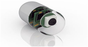
CAPSULE ENDOSCOPY:
IMAGE OF CAPSULE ENDOSCOPE
IMAGE OF CAPSULE ENDOSCOPY INSIDE THE STOMACH
Capsule endoscopy is a procedure that uses a tiny wireless camera to take pictures of your digestive tract.
A capsule endoscopy camera sits inside a vitamin-size capsule you swallow.
As the capsule travels through your digestive tract, the camera takes thousands of pictures that are transmitted to a recorder you wear a belt around your waist.
It also helps to view the area that isn’t easily reached
It is mostly done when the cause of gastrointestinal bleeding, cancer diagnosis, polyps screening etc...
RISK AND SIDE EFFECTS OF ENDOSCOPY:
Reaction to sedation prior to your upper endoscopy some sedation has to be given after the procedure, sometimes it causes the adverse risk of a serious reaction.
Sometimes tearing of the gastrointestinal tract may occur.
Mostly endoscopies consist of an examination and biopsy and so the risk of infection is low. When the risk of infection increases when additional procedures are performed.
If the biopsy has been done then the risk of bleeding complication may occur. In rare cases, such bleeding may require a blood transfusion.
SIGNS AND SYMPTOMS AFTER ENDOSCOPY:
Vomiting
Fever
Chest pain
Difficulty in swallowing
Shortness of breath
Persistent abdominal pain
Bloating and gas
Cramping
Sore throat
ABNORMALITIES DETECTED DURING ENDOSCOPY:
Mucosal abnormalities are detected such as erythema, ulcer, erosion and polyp.
Narrowed areas and strictures of the oesophagus, stomach or duodenum are detected and treated.
Objects stuck in the oesophagus or stomach can be detected and removed.
Bleeding due to ulcers can be detected.
Precancerous abnormalities such as Barrett’s oesophagus is detected.
Inflammation or swelling can be detected.
Blockages are detected.
Gastroesophageal reflux disease ( it is a digestive disease in which stomach acid or bile irritates food pipe) is detected, for this biopsies are needed to diagnose.
HOW TO MAINTAIN ENDOSCOPE:
Maintenance is very important for medical equipment.
Pre-cleaning is important to clean priorly for the disinfection after the procedure done immediately wipe the endoscope using an approved enzymatic solution. Wash suction, air and water channels according to the manufacturer’s instruction.
Always test the endoscope for leaks, immerse in water and test.
Thoroughly rinse the exterior of the endoscope using clean water.
Clean all channel with proper disinfectant.
Use alcohol-water mixture (70 to 80 percent ) to flush all, then wipe and dry.
Store the endoscope in a well-ventilated container.
TROUBLES IN ENDOSCOPE:
The left-hand holds the control section with the right hand controlling the insertion tip, be aware of kinking of the upper end of the umbilical cord. If it changes during bending it could cause damage in the internal channel.
The most proximal portion of the endoscope is the eyepiece. The generic replacement eyepiece commonly repairs by unauthorized repair firms in which they use low-quality materials so during the examination it may cause an undesirable view.
Sometimes the shaft (it contains all illumination components) in the endoscopes may get damaged.
HOW TO TROUBLESHOOT THE PROBLEMS:
Correct positioning for each procedure results in the safest operation/endoscopy.
The high-quality standard authorized eyepiece is fitted according to the manufacturer’s specification.
Always use the endoscopes with proper guidance.
👉Please Watch Our Endoscope (Part 2) Video From Our YouTube Channel Below:-
An endoscope is a thin, rigid, flexible instrument combined with fibre-optics and charge-coupled device to facilitate illumination or visualization of the interior organs. Endoscopy is a process in which the endoscope is passed through the mouth, into the oesophagus and down towards the stomach and duodenum. The tip of the endoscope contains the light and tiny video camera so that the doctor or operator can able to see inside your gut.
The endoscope has a channel through which surgeons can manipulate tiny instruments, such as forceps, surgical scissors, and suction devices.
A variety of instruments can be fitted to the endoscope for different purposes.
Through one channel of endoscope water and the air is conducted to wash and dry the surgical site.
Endoscope gives visual evidence of the problem such as ulceration or inflammation.
HISTORY OF THE ENDOSCOPE:
In 1805, Philip Bozzini made the first attempt to observe the living human body directly through a tube, he created lichtleiter a light-guiding instrument.
IMAGE OF PHILIP BOZZINI AND LICHTLEITER ENDOSCOPE
In 1853, Antonie Jean Desormeaux of France specially designed to examine the urinary tract and the bladder. He named it as an endoscope.
IMAGE OF ANTONIE JEAN DESORMEAUX ENDOSCOPE
In 1868, Dr. Adolph Kussmaul of Germany succeeded in taking a look inside the stomach of a living human body.
IMAGE OF Dr.ADOLPH KUSSMAUL ENDOSCOPE
In 1932, Johann von Mikulicz and his associates created the 1st rigid gastroscope but it is not flexible.
In 1932, Dr. Rudolph Schindler invented a flexible gastroscope that allows the examinations even while the tube is bent. This tube was 75 centimetres in length of the tube and 11 millimetres in diameter.
IMAGE OF Dr. RUDOLPH SCHINER FLEXIBLE GASTROSCOPE
In 1983 the first endoscope without fibre optic transmission of the image was produced by Welch Alleyn Incorporated in New York. At the tip of the instrument was an electronic sensor consisting of a packed grid of photocell receptors which transmitted images electronically to a video processor and then to a television monitor.
IMAGE OF WELCH ALLEYN ENDOSCOPE
WHY ENDOSCOPY IS NEEDED?
The doctor may recommend an endoscopy for various reasons
If the food pipe (oesophagus), stomach, or top part of the small intestine need to be looked at, then gastroscopy is needed.
Endoscopy is needed for the early detection of cancer from the mucous covering in either the upper or lower tracts of the digestive tube.
To find out helicobacter pylori infections, it is the bacteria are thought to cause gastric tumours.
Gastroscopy is needed for the persons with gastroesophageal reflux disease or GERD, especially for the person, those who consume alcohol and smoke regularly and complain of chronic heartburn are at risk for cancer of the oesophagus.
It is needed to find out the changes in the lining of your oesophagus.
IS THAT PREPARATION NEEDED FOR ENDOSCOPY?
Endoscopy (gastroscopy) requires special preparations:
It is advisable to discontinue taking alcoholic beverages and also avoid nuts, seeds and spicy foods before the examination.
The last meal before the diagnostic procedure must be no later than at 6 PM of the previous day.
On the day of the examination, it is not allowed to take foods or beverages for 6 to 8 hours before the procedure, a little water can be taken no later 2-4 hours before the examination.
If the patient takes any chronic medication in caps or pills such as any medicine that used to treat diabetics, such as insulin and metformin, any blood thinning medication ( i.e) low-dose aspirin, aspirin, warfarin or clopidogrel, non-steroidal anti-inflammatory medicine, they should suspend taking them since foreign items in the cavity of the organ being examined can distort the picture.
It is very important to alert the doctor about any existing allergic reactions if they are associated with medical drugs.
Before the examination the doctor will administer premedication or anesthetization of the base of tongue and throat with a spray, this will reduce discomfort and pain.
If the patient has any heart valve disease and has pacemaker priorly inform to the concerned doctor.
I suppose the patient may wear dentures they may be asked to remove them prior to their procedure.
Patients are advised to leave their contact lenses at home and wear glasses instead.
IS THAT ANY PROCEDURE IS THERE DURING ENDOSCOPY (GASTROSCOPY)?
The patient will be asked to lie flat, generally on the left side, before the procedure, your throat will be numbed with a local anaesthetic spray, you can choose to have sedative if you prefer. This means you will still be awake but will be drowsy and have reduced awareness about what’s happening.
A smallmouth guard will put between our teeth to stop you from biting the endoscope and to protect the teeth.
The endoscope will be placed into your mouth and you will be instructed to swallow it down to oesophagus and into your stomach.
The doctor will direct air into your stomach via the gastroscope, this will make viewing easier.
Sometimes a special instrument can be inserted through the scope, and a small sample of tissue removed (biopsy) This is not painful.
Some treatments can be performed while the endoscope is in such as removing polyps, controlling blood loss from an ulcer, controlling or preventing bleeding from enlarged veins, a narrowed oesophagus can be widened.
COMPONENTS OF ENODOSCOPE:
A thin, long flexible, rigid tube
A lens or lens system
A light-transmitting system
The eyepiece
Control system
Water pipes
The operational channel
Control cables.
A THIN, FLEXIBLE, RIGID TUBE:
FLEXIBLE: Flexible endoscope allows the user to navigate hard to reach areas by controlling the directional movement of the scopes distal end.
IMAGE OF FLEXIBLE TUBE
RIGID: Rigid endoscopes, they are small tubular telescopes that allow the physician to look inside joints and body cavities that otherwise could only be examined through a non-invasive procedure.
IMAGE OF RIGID TUBE
A LENS OR LENS SYSTEM:
IMAGE OF LENS
A lens to transmit the image of the patient’ internal system to the operator or viewer ( this is generally a relay lens in rigid endoscopes or multiple fibre-optics for fiberscopes), object lens allows visualization of mucosa.
A LIGHT TRANSMITTING SYSTEM:
A system to transmit light to enhance the visibility of the area of being examined ( the source of this light is usually based outside of the body, directed through optical fibres).
THE EYEPIECE:
IMAGE OF EYEPIECE
An eyepiece in video scopes lacking eyepieces, images from inside the patient is sent to a screen for viewing and capture.
WATER PIPES:
IMAGE OF WATER PIPES
The pipes serve to carry water which is used to wash the lens thereby maintaining a clear view.
THE OPERATIONAL CHANNEL:
IMAGE OF OPERATIONAL CHANNEL
This is an opening of the device that is used to move various accessories to the distal end (of the endoscope) for surgery purpose, for example, biopsy channel this allows passage of biopsy forceps and other instruments to undertake therapy.
CONTROL CABLES:
IMAGE OF THE CONTROL CABLES
This is used to control the direction
that the distilled end will bend as it moves through body cavities.
TOOLS USED WITH ENDOSCOPE:
An endoscope has a channel through
which the doctor can insert tools. These tools collect tissue or provide
treatment. Types of tools are:
FLEXIBLE FORCEPS: These tong-like tools take a tissue sample.
IMAGE
OF FLEXIBLE FORCEPS
BIOPSY FORCEPS: These remove a tissue sample or a suspicious growth.
IMAGE OF BIOPSY FORCEPS
CYTOLOGY BRUSHES: These take cell samples.
IMAGE OF CYTOLOGY BRUSHES
SUTURE REMOVAL FORCEPS: These remove stitches inside the body.
IMAGE OF SUTURE REMOVAL FORCEPS
👉Please Watch Our Endoscope (Part 1) Video From Our YouTube Channel Below:-
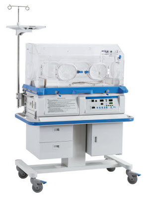
Infant incubator or isolette is a biomedical device in which it provides warmth, humidity and oxygen in a controlled manner as required by the newborn baby and also it maintains a healthy and clean environment.
HISTORY BEHIND INFANT INCUBATORS:
During 1891 the modern incubator is invented by DR.Alexandar Lyon.
In 1907 Pierre Constant Budin proposed the study of the influence of body temperature on infant mortality.
In 1932 Julius Hess in his patents for incubators proposed a mechanism of addition of oxygen supplement in the incubator.
Later 1933 Blackfan and Yaglaw released the report for the improved survival on newborn infants nurtured in humidity enriched environment.
DIAGRAMATIC REPRESENTATION OF THE PARTS OF INCUBATOR:
DEPARTMENTS THAT USE INCUBATORS:
NICU (Neonatal Intensive Care Unit)
SCN (Special Care Nursery)
Postnatal care wards
Labour ward(sometimes transport incubators are found to transport the newborn to NICU.
SCIENCE BEHIND INFANT INCUBATORS:
Generally, infant incubator is used to provide needed temperature for a baby, so the thermoregulation process is incorporated with it.
WHAT IS THERMOREGULATION?
Thermoregulation is the ability of the body to equalize the heat produced and heat lost by the body thereby maintain the body temperature in the normal range. The normal body temperature is 37°C which this temperature is maintained by the hypothalamus. If the skin temperature drops below 37°C, a variety of responses are initiated to conserve the heat in the body and to increase heat production. Heat may be produced in different activities that taking place in our body mainly during metabolic activity. Heat lost may occur mainly by these categories they are Evaporation, Radiation, Conduction and Convection.
SCHEMATIC REPRESENTATION OF THE PROCESS OF INFANT INCUBATOR
THERMOREGULATION PROCESS IN UTERUS:
Essentially the baby in a coldblooded creature so it unable to thermoregulate like adults, therefore, it adapts the mother core temperature, Generally the baby’s core temperature is always slightly 0.5°C above than the mother core temperature. When this thermal instability causes the baby cannot able to breathe and some risk has arrived. Even this instability can last for several days or even weeks.
WHY INCUBATORS IS NEEDED:-
WHY PREMATURE NEONATES NEED INCUBATOR:
Commonly babies are born more than 37 weeks if the babies are born less than 37 weeks the babies are admitted in NICU(Neonatal intensive care unit). In NICU the premature baby is quite physically and developmentally developed and those are unable to transition to the normal outside environment. They can’t control their body temperature and also it shows many unstable vital conditions, So these babies need to remain in the controlled environment known as incubator it is also known as the isolette. In which it provides heat to keep constant body temperature and also maintain the uterine environment.
WHY WE NEED INCUBATORS:
The incubators are used to provide a constant temperature. In NICU the bright light causes a negative impact on the infants in which the infants are so sensitive and fragile bodies. Incubators reduce direct light exposure thereby it helps the infant to reduce strain. Other than the temperature it helps to provide the proper oxygen supply to the infants.
PRINCIPLE OF INFANT INCUBATORS:
In incubators underneath the baby are an air-blown electric heating system and humidification system in which it circulates heated humid air at a desired temperature and humidity through the incubator chamber. Oxygenation also provided for the infant.
WORKING PRINCIPLE:
The mattress is present in the incubator where the baby lies is completely enclosed by a clear plastic canopy.
In this temperature and humidity are the parameters which have to be controlled.
A temperature sensor is tapped into the baby skin and the incubator heater adjusts to maintain the baby at a constant temperature.
The isolation chamber that helps to regulate the temperature of an infant and can provide air which is enriched in humidity/oxygen.
The temperature in the incubator is increased or decreased by a heating element which is placed below the mattress.
PREPARATION OF THE INFANT INCUBATOR:
Pre-warmed the desired temperature based on the infant age, size and condition.
Use air mode and must once calibrate the motor running if in use for a baby.
Check and record the temperature, humidity and oxygen levels.
Ensure that alarms are working automatically.
No equipment is placed in the top of the canopy.
Re-check the temperature adjustments.
Before using the infant incubator it must be cleaned and sterilized.
FUNCTION OF INFANT INCUBATOR:
Shielding infants from germs and microorganisms.
Temperature control.
Humidity control.
Breathing gas filtration.
O2 concentration.
TYPES OF INCUBATORS:-
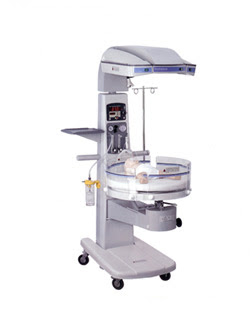
1) OPEN BOX INCUBATORS: This open box incubator is also known as Armstrong incubators, provide radiant heat below the baby but are otherwise open to the air, allowing for easy access.
2) CLOSED BOX INCUBATORS: It has a fresh air filtration system that minimizes the risk of infection and prevents the loss of moisture from the air.
3) DOUBLE-WALLED INCUBATORS: It has two walls that can further prevent heat and air moisture loss.
4) SERVO-CONTROL INCUBATORS: These are automatically programmed to adjust temperature and humidity levels based on skin sensors attached to the baby.
5) PORTABLE INCUBATORS: This is also known as transport incubators, are used to move the newborn from one part of the hospital to another.
BLOCK DIAGRAM OF INFANT INCUBATOR:
BLOCK DIAGRAM OF TEMPERATURE AND HUMIDITY CONTROLLER
BLOCK DIAGRAM OF THE POWER SUPPLY UNIT
COMPONENTS OF INFANT INCUBATOR:
HEATER:The room air that is passed over the heater is heated to the desired temperature which the infant is needed.
HUMIDIFIER: The heated air pass through the humidifier to be humidified to the desired range.
FRONT PANEL: This is used to interface with the machine to get the desired operation.
FILTER: The filter that is used in the incubator is to protect the infant from airborne bacteria and also the impurities /germs.
BLOWER: The fan/blower is used to circulate the air in the infant incubator.
POWER SUPPLY: The power supply provides the necessary voltage required by the infant incubator.
ADVANCEMENTS IN INFANT INCUBATORS:
Since the 19th century, the devices termed as incubators were developed to maintain thermal stability for underweight newborn and sick babies thus improving the chances of survival. Many advancements are developed for infant incubators such as mechanical ventilators specially designed for neonates. There are many parameters included in infant incubators such as oxygenation support, ECG for low heart rate babies and continuous predictive monitoring are available.
RISK IN INFANT INCUBATOR:
Mechanical stress has damaged temperature. The control mechanism, in which in turn have overheated infants, causing brain damage or death.
High noise level causes the risk of newborn babies.
Illumination of light causes the great risk of the visual pathway of the newborn.
Sometimes due to lack/more of oxygen may lead to shallow respiratory problems such as APNEA(irregular respiratory pattern).
Hypothermia is caused this is characterized by lowering of the body temperature than 36°C.
TROUBLES ARISES IN INFANT INCUBATOR:
Lack of maintenance results in the use of incubator with high-ambient noise levels which cause misaligned air-circulating fan results in hearing loss.
The defective door may sometimes cause damage.
Sometimes there is damage in the chord arise problems.
HOW TO TROUBLESHOOT THE PROBLEMS:
Servo-control incubators are automatically programmed to adjust temperature and humidity based on skin sensors that padded on the newborn babies.
Open-box system provides radiant heat below the baby easy access to outside.
PREVENTIVE MAINTENANCE OF INFANT INCUBATOR:
Be sure that the plastic housings are intact, that all hardware present and tight, and serious abuse. Remove any tape adhere to the unit, check all rubber and plastic gaskets in the unit.
Verify all parts are assembled correctly. The misassembly of internal components that could interfere with performance.
If the device is mounted on a stand or cart, examine the condition of the mount.
Check all the nuts and bolts ate tightened fully. Use the screwdriver.
Examine the AC power plug for the damage. If damaged replace the entire cord.
Check the condition of all tubing, cuff, and bulbs.
Examine all fittings and electrical cable connectors for general condition.
Disassemble the heating unit enough to expose the heating element.
Inspect the fan blades for deterioration and damage.
Inspect the physical conditions of the battery and battery connectors.
Since incubators carry oxygen, a fire hazard sign may be visible.
Check the action of the primary and safety thermostats with the fully assembled.
Clean the exterior and interior.
Calibrate if needed.
ROLE OF BIOMEDICAL ENGINEERS:
As a biomedical engineer, we have to maintain the equipment such as calibrate the equipment for a regular period of times, check the power sources before use, the above preventive maintenance has to be checked by every biomedical engineer.
👉Please Watch Our Infant Incubator Video from Our YouTube Channel Below:-