Post Top Ad

WE DO ADVERTISEMENT SERVICES FOR YOUR HEALTHCARE PRODUCTS, PROMOTING YOUR HEALTHCARE EVENTS, TECHNICAL REVIEWS FOR YOUR MEDICAL DEVICES, ETC...
CONTACT US 📞 +94 76 911 1820 FOR FURTHER DETAILS ABOUT OUR SUPPORTS.....
OUR EMAIL ADDRESS:- sam.gastondiaz@gmail.com
Advertise with Learn BioMed Engine
Promote healthcare products, events, training programs, and medical technology services.
Email: healthcareengineeringteam@gmail.com | WhatsApp: +94 76 911 1820

Friday, September 17, 2021

Cryotherapy

Introduction:
Cryo-therapy is also known as Cryo-surgery or Ice-surgery. It uses cold temperature to damage tissue to treat disease.

What is Cryotherapy?
  • Cryotherapy means it’s the process of using high or extremely cold temperatures in order to destroy or damage the tissue by freezing those.
  • It helps to treat various skin diseases and also some cancer.
  • Substance like liquid nitrogen is used by doctors.
  • Cryotherapy reduces muscle pain, swelling twist in ankles or in wrists.

Principles of Cryotherapy

Lewis Hunting Reaction:
“The process of alternating vasodilation and vasoconstriction in extreme exposure to cold is called the Lewis Hunting Reaction”

At first, vasoconstriction occurs first to reduce extremities. This is the result of the sudden decrease in the release of neurotransmitters due to local cold from the sympathetic nerves to the muscular coat of the arteriovenous anastomoses. This increases blood flow and the temperature of the fingers. Vasodilation is followed by the new phase of vasoconstriction after that the process repeats itself.

When cold is maintained for more than 15 minutes or reduced below 10 degrees Celcius, vasodilation is cold-induced after the initial time of vasoconstriction.

Cryobiology :
Cells of mammals can be destroyed by cooling to the temperature of -20 degrees Celcius. Information of ice crystals of both intracellular and extracellular begins the primary injury. The outer membrane of the cells ruptures by intracellular crystals and ice formation outside the cell which dehydrates the cellular environment which results in lethal electrolyte concentrations and changes in pH. The time cell organelles are damaged, the cell loses the ability to regulate ions which ensures cell death.

Even though liquid nitrogen, nitrous oxide, and carbon dioxide are all cryogens used in many medicines. Liquid nitrogen is the most versatile. Hence it is most commonly used. The boiling point of liquid nitrogen is -195.8 degrees Celsius. The liquid form of cryogens stored in Dewar flasks can be delivered as a spray. 

Mechanism of Action:
Cryotherapy can be also known as cryosurgery. It is commonly used to treat a variety of Benign and Malignant Lesions. In Cryotherapy the mechanism of destruction is necrosis. Necrosis is the result of the freezing and Thawing of cells. Tissue injury begins with prolonged tissue cooling, metabolic disruption, ice crystal formation, and cellular rupture. Microcirculatory failure and Ischemia lead to cell death, resulting in coagulative necrosis after having.

Physical destruction procedures are effective immediately, but physiological effects which include induction of apoptosis and cytokine release and secondary necrosis produce damaging effects over several days.

The mechanism of action in cryotherapy is divided into 3 Phase as below:-
  • Heat transfer.
  • Cell Injury.
  • Inflammation.
a) Heat Transfer:- 
                There is a mechanism by which cryotherapy destroys the targeted cells. The mechanism involves the quick transfer of heat from the skin to a heat sink. Liquid nitrogen is the most commonly used cryogen, which has a boiling point of 196 degrees celsius. The rate of heat transfer depends on the temperature difference between the skin and the liquid nitrogen. In the spray cryotherapy technique, the liquid nitrogen is directly applied to the skin and the heat in the skin is transferred to the liquid nitrogen. This process results immediately. If a cryoprobe is used in the cryotherapy conduction heat transfer occurs in which the heat is transferred via the copper-metal probe.

b) Cell injury:-
    Once the cell is frozen, cell injury occurs during the thaw. Ice crystals do not form until the temperature reaches –5 to -10 degrees Celsius because of hyperosmotic intracellular conditions. The water to ice transformation concentrates the extracellular solutes and leads to an osmotic gradient across the cell membrane, leads to further damage. 

The tissue of epithelial cells damage maximum with rapid freezing and slow thaw and are suitable for the malignancies treatment. After rapid thaw fibroblasts produce less collagen. Hence a rapid thaw is much more suitable for the treatment of keloids or benign lesions. When a steak is defrosted from the freezer freeze damage occurs.

When fully thawed the steak juices come out which represents that the intracellular liquid that has escaped because of the damage to the cell wall. By concentrating electrolytes intracellularly low temperature also ensures maximum damage. Benign lesions need a temperature of -20 to -25 degrees Celsius, whereas malignant skin cancers require a temperature of -50 degrees Celsius.

c) Inflammation:-
   Inflammation is the last response to cryotherapy which is seen as erythema and edema. Inflammation helps in cell destruction and is the response to cell death. Cryotherapy treatment causes separation of basement membrane and which may also result in blister formation.


Procedure Performed During The Cryotherapy:
Cryotherapy is performed by a well-trained radiologist in a radiology suite or in the operating room. It is an image-guided procedure. On the procedure table patient will be positioned and it is an outpatient basis procedure. Doctors will apply liquid nitrogen to the area with a spray device or cotton swab if cryotherapy is done topically.

 A percutaneous procedure is performed for tumors that are deep inside the body and can be approached through the skin. A thin, needle–size applicator or cryoprobes are inserted.


  • Monitors are used to tracking blood pressure, heart rate, oxygen level, and pulse. 
  • The patient will be connected to the monitors. 
  • An intravenous(lV) line is inserted into a vein in the arm or hand of the patient by a technologist or nurse to administer a sedative. 
  • Breathing tubes may not be required in this process. 
  • General anesthesia is required in some patients may be. 
  • The surface will be shaved, sterilized, and covered with a sterile drape where the applicators or cryoprobes are to be inserted. 
  • A small skin incision is made at the site.
  • Through the guidance of imaging, applicators or cryoprobes will be inserted through the skin to the site of the diseased tissue. 
  • Argon gas or liquid nitrogen is delivered once the applicators are in place. 
  • A rapid decrease in the temperature at the tip of the probe leads to the creation of an “ice ball”. 
  • Due to this all the water around the tip of the probe freezes. 
  • Ultrasound, CT, or MRI are used to visualize ice balls. 
  • lV line and applicator are removed. 
  • Application of pressure stops the bleeding and skin will be covered with a bandage. 
  • Here sutures are not required. 

Types of Cryotherapy


1. Ice Packs:- It is the most common method of cryotherapy. There are different types of ice used in ice packs. The most common types are ice packs made with cubed, crushed, and wetted ice.  It was discovered that wetted ice is better to lower surface temperature during treatment and maintaining the lower temperature during recovery. It is also more effective in lowering the intramuscular temperature during treatment.

2. Ice Spray:-  A cooling effect can also be produced by icing spray for a similar effect.

3. Immersion:- Simple or whirlpool immersion.

4. Ice Massage:- slow strokes in a circular motion for  5-10 mins.

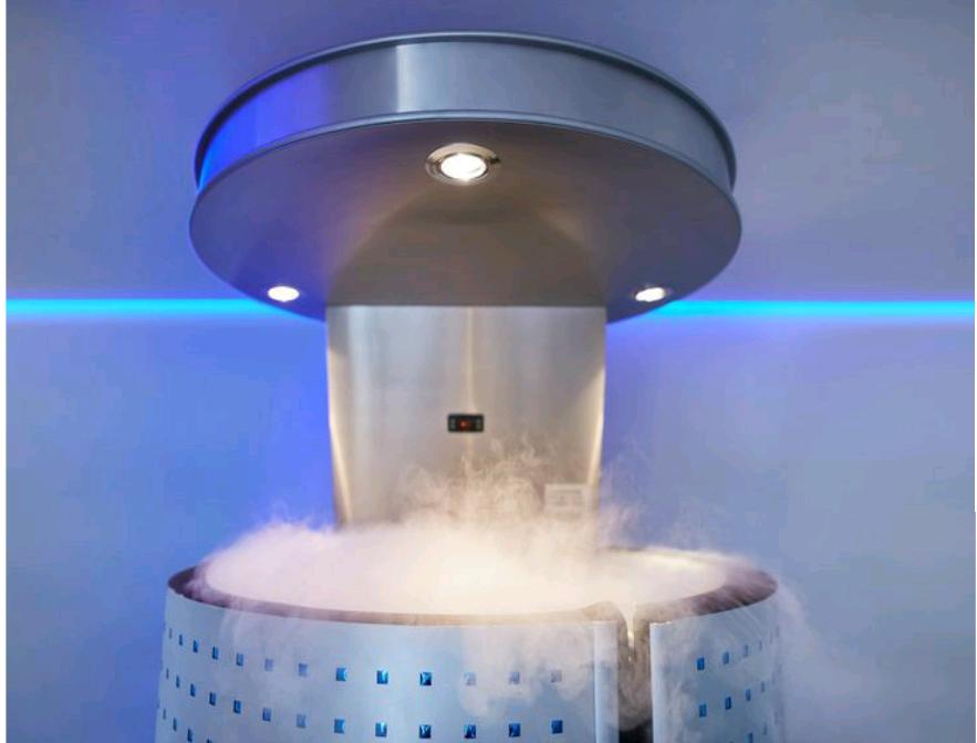
More recently whole body cryotherapy has become popular for athletes, to help aid recovery, as well as in persistent pain patients such as rheumatological conditions. More research is needed to understand the effect on the body and its relation to pain.


Benefits Of Cryotherapy
1. Helps to reduce migraine:- Cryotherapy helps in treating migraines as it cools and numbs the nerves around the neck area. By applying a neck wrap containing 2 frozen ice packs to the carotid arteries reduces migraine pain. The carotid arteries are accessible and also they are close to the carotid arteries.

2. Irritation of Numbs nerve:- Cryotherapy has been using to treat injuries of athletes. The cold can actually numb an irritated nerve.

3. Helps to treat mood disorder:-  The ultra-cold temperature in whole-body cryotherapy can cause It includes the release of adrenaline, noradrenaline, and endorphins. It was actually effective in short-term treatment.

4. Reduces arthritic pain:-  The whole–body cryotherapy significantly reduced pain in people with arthritis. As a result, it has also allowed for physiotherapy and occupational therapy.

5. May help treats low-rise tumors:-  It can be used as a cancer treatment.  It is done by freezing cancer cells and surrounding them with the help of ice crystals. It is used to treat some low-risk tumors.

6. May also help to prevent Alzheimer's disease and dementia:-  It may be an effective treatment because the anti-oxidative and anti-inflammatory effects of cryotherapy could help combat the inflammatory and oxidative stress responses that occur with Alzheimer's.


Advantages Of Cryotherapy:
  • It can temporarily reduce nerve activity, which can also relieve pain.
  • This therapy can treat tissue externally and internally.
  • It can be used to treat a variety of skin conditions and some cancers.

Disadvantages Of Cryotherapy:
  • Cramping around the cervix after cryotherapy and bleeding.
  • Skin infection, scarring, and swelling.
  • Bone fractures
  • Headache, hair loss, and hypopigmentation.


Article Contributors:-


Friday, September 10, 2021

Fluoroscope

 INTRODUCTION 

What is Fluoroscopy?

Fluoroscopy is a medical imaging procedure that uses an x-ray beam that goes continuously through the body to make an image and it is also a sort of x-ray that displays organs, tissues, or other interior structures moving in real-time. The image is projected on a monitor, allowing the doctor to watch the inside organs move in real-time.

Fluoroscopy is used to aid in the diagnosis of abnormalities with your,
1. Esophagus
2. Stomach 
3. Duodenum
4. Small bowels

Doctors can use this approach to see the flow of oral contrast through the digestive system in real-time.
The fluoroscopy examination will scan your,
1. Upper GI
2. Small bowel

Inform the nurse or technologist if you are allergic to any medications or if you have recently undergone a high-density contrast procedure. You may take your normal medication with modest amounts of water the day before and after the exam. The stomach and small intestine will be shown in the small bowel series. You should not undergo any contrast studies within 24 to 48 hours of this exam.


Fluoroscopy Machine:
 
An endless stream of x-ray images. Around 25-30 images per second. Images are shown on a computer monitor. Similar to a television screen.


HISTORY OF FLUOROSCOPY.

Roentgen's X-Ray: 

1895 Thomas Edison exposed over 8000 chemicals four months later. Calcium tungstate has the most fluorescence and was used to make the Edison Vitascope (device for recreational Fluoroscopy). After the death of his assistant, Thomas never returned to X-Ray research. He examined his hand with a custom-made fluoroscope. Clarence Dally, his aide, died as a result of radiation. 

Early fluoroscopes were basically cardboard funnels, open at the narrow end of the observer and closed with a skinny cardboard piece coated on the inside with a film of fluorescent metal salt. The fluorescent screen produces only a small limited light. The fluoroscope image was faint, so the radiologist went into the darkroom to adjust their eyes to the darkness. William Trendelenburg created red adaptation goggles. 

Russel H. Morgan invented the image intensification tube in the 1940s. Image visibility is improved, the radiation dose is reduced, and a persistent image is recorded out of light.


How It Works?

The machine utilized in fluoroscopy is called fluoroscope. The fluoroscope is a sort of x-beam machine that can either utilize a beating or steady x-beam bar. The x-beam machine has an x-beam tube that is either made of glass or metal and has a vacuum seal inside. It produces an x-beam by changing over power from its electrical cable. The electrical cable has a flow of 120-480 volts, anyway it should be changed over into a 25-150 kilovolt range. 

Then, at that point, it creates a current of electrons that are taken shots at a tungsten target. At the point when the electrons strike this objective, the electrons stop and make the arrival of the body being imaged. These electromagnetic waves can go through the body and make images of the inside structures. Various tissues in the body have various densities; along these lines, the tissues that are less thick ingest less of the x-beam. 

The diverse in the measure of waves being assimilated is the thing that makes a distinction in the openness and permits the image to be more definite. With the fluoroscope, when the pillar goes through the body, it hits an image intensifier, which builds the splendor of the picture, so it very well may be seen on a presentation screen. 

The picture intensifier likewise contains a camcorder that catches the two-dimensional examples of light as a video from the x-beam machine. Then, at that point, the sign is changed over once more into an example of light, which can be seen on the screen, as an image Illustrate the parts of an analog fluoroscopy image intensifier tube.


There is an input phosphor there is a photocathode right behind the phosphor. Then there are series of electrostatic lenses that push electrons towards a focal point & this accelerating anode towards. Which the electrons are being attracted and finally there is an output phosphor. 

So, what we are doing is we are converting the x- rays ate coming into the fluoroscopy. They are hitting this input phosphor and the input phosphor is glowing. It's giving off light x- rays hit the phosphor and the glow right. Then this photo Catherine picks up the light and turns that into electrons. Right turns the light into electrons which are then accelerated towards this anode. 

Very similar to what's happening inside of an x-ray tube right. We had the production of electrons they are accelerated towards an anode in this case. They are not going to strike the image instead. 


The Physics of Image Intensifier that which Used in Fluoroscopy:

Originally the conventional fluoroscopy way back when they didn't have a way to do that so the images that they were looking at were very dim and they had to use what are called rods in the eye & the rods see dim objects. They see black & whites and so the whole purpose of bringing the brightness up the brightness level up is to change the vision into the co vision which have they see much better & we'll get into a little bit more of that just a minute. So, a bottom line is they were developed to increase the brightness & they increase it a great deal 


We are just increasing the brightness exponentially over the original image, this particular image shows us the entire system, and also on the side, you're going to see an eye - without casing on its side.

The eye itself and in this particular image, you can see the x-ray tube below the table. The beam interacting with the patient & the remnant beam coming into the image intensifier itself. 

The remnant beam radiation coming from the patient interacts with the input phosphor and at the input phosphor radiation. The information it carries is changed into light photons and then the light photons hit the photocathode where they're changed into electrons & they are accelerated at a very high speed as well as being compressed by the electrostatic lenses which are following the size of the periphery of the image intensifier tube. 

They come to a focal point and at that point, they actually cross over, and then they come out of a very small output phosphor it's about one inch. You have a very large curve for the input phosphor & the very small output phosphor. 

This whole process is what gives us that extreme brightness that we have in our image and then once it comes out of the output phosphor there are some things that happen to it.

Flux Gain: Measurement of the input light photons due to the conversion efficiency of the output screen.

Brightness = Flux Gain X Magnification Gain


Fluoroscopy Quality Control:

Exposure rate:-
  • Normal fluoro: ESE < 10 R / Min 
  • Interventional fluoro: ESE < 20 R / Min
  • Cineradiography: Unlimited exposure rates. 

Spot film exposure:-
  • Cassette ESE approximately 200mR per spot.
  • Photo fluoro spot ESE approximately 100mR per spot.

Automatic brightness control (ABC):-
  • Evaluate annually. 

Patient dose during fluoro: Conventional vs Digital 

            Patient dose

            Conventional

            Digital

5-minute fluoro

200mGy (20 rad)

100mGy (10 rad)

3 spot films - normal mode

6mGy (0.6 rad)

2mGy (0.2 rad)

3 spot films – mag mode

10mGy (1.0 rad)

3mGy (0.3 rad)

Total Dose

216mGy (2.16 rad)

105mGy (10.5 rad)


Image Intensification

Input phosphor:


  • Mad of cesium iodide (CsI)
  • Receives radiation exiting patient. 
  • Emits light photons.
Photocathode:
  • Responds to the light exiting input phosphor. 
  • Emits electrons. 
Electrostatic lenses:
  • Focus electrons. 
Output phosphor:
  • Receives electrons from the photocathode. 
  • Emits 50 – 75 × more than received by the photocathode

Distortion.
As the size of the input phosphor is usually very large as the size of the input phosphor is increased or decreased. You're increasing and decreasing just the Distortions. If you have left it wide open say 25cm is your largest size if you leave it at 25. You will see in your image is quite a bit of distortion in the periphery.

1. Pincushion distortion:
    Caused by projecting an image formed on a curved input phosphor to a flat output phosphor.

2. Vignetting:
    Decrease in brightness or light intensity- periphery of the image.


3. Veiling Clare:
  •     Scattered light in the output window 
  •     Increase your background signal &it decreases the actual contrast of your image. 

Charge Couple Device:- 

Advantage of the charge-coupled device for medical imaging. 
  • High spatial resolution. 
  • High signal to noise ratio. 
  • High detective quantum efficiency (DQE).
  • No warm-up is required. 
  • No leg or blooming. 
  • Lower patient dos. 
  • Unlimited life. 
  • No spatial distortion. 

Creating a Fluoroscopy Image:-

The amount of radiation required varies and it’s based on the procedure. A necessary characteristic of Fluoroscopy is sensitivity. Amount of exposure needed to form an image. Non-intensified Fluoroscopy utilizes a fluorescent screen only just for a receptor. Key facts
  • Generally real-time exams
  • Typically uses an under-table tube and radiation is shooting upward
  • Exam dependent
    • May needs the use of contrast media 
    • X-ray altering media that either positively or negatively affects the image counting on what is used to make something stand.
  • X-ray tube & imaging device mounted on C-arms to keep up SID

PROCEDURE OF FLUOROSCOPY 

What happens during a fluoroscopy procedure?
  • We will be asked to remove any clothing or jewelry that may interfere with the procedure, and depending on the type of procedure, we may be asked to: assume different positions, move a specific body part, or hold our breath at intervals while the fluoroscopy is performed.
  • A special x-ray machine will produce fluoroscopy views of the body structure being examined or treated.


CONTRAST PROCEDURE
  • During the test, you will be asked to change into scrubs or a hospital gown. Your personal item will be secured in a looker you will stand between a table and a fluoroscopy machine. 
  • You will drink contrast medium. You will be asked to turn in different positions. This will allow the camera to take images of your abdomen from all angles. 
  • Multiple peeping sounds can be heard during the exam.
    • Upper GI exams will take approximately 30 minutes. 
    • Small bowel series exams will take 1 to 4 hours longer. 
  • After the test, you will be discharged. Immediately after the exam, there are no driving restrictions. Drink at least 6 to 8 ounces of fluids preferably water. You may eat a regular diet include Fiber unless otherwise instructed to do so by your doctor. There is no radiation to concerned with your doctors will discuss the results with you at your next appointment 


Barium X-Rays procedure 
  • Fluoroscopy used alone.
  • Give the physician the opportunity to check movement in the intestines.
  • Barium moves through them during the procedure.
  • Aids physicians in inserting a catheter.
  • Likewise helps them in identifying blockages in arteries.
  • Physicians can see the flow of blood.

Overall Procedure of Fluoroscopy
  • Inclusion of an IV into the patient’s arm or hand.
  • The patient moved onto the X-Ray table.
  • Additional lines could also be inserted for catheter procedures.
  • X-Ray scanner helps to make Fluoroscopic images of the body.
  • Dye could also be injected into the IV at this point.
  • The type of care is going to be selected after the procedure has finished.

What is it used for:-
Fluoroscopy and other medical imaging procedures serve an important role in avoiding health problems and identifying diseases. Our doctor may request that we undergo fluoroscopy during a hospital stay or outpatient procedure to determine treatment options for a specific health condition.

Fluoroscopy is a technique that is utilized in a variety of imaging treatments. The following are some of the most prevalent fluoroscopy applications:
  • Barium swallow or Barium enema:- Fluoroscopy is used to show the movement of the gastrointestinal (digestive) system during various procedures. 
  • Cardiac catheterization:- Fluoroscopy is used to visualize blood flow through the arteries, during this treatment. It's used to diagnose and treat some heart problems. 
  • Placement of catheter or stent inside the body:- Catheters are hollow, thin tubes. They are used to get fluids into the body or to remove excess fluids. Stents are medical devices that help unblocked blocked or narrowed blood arteries. Fluoroscopy is used to check that these devices are properly placed. 
  • Hysterosalpingogram:- Fluoroscopy is utilized to produce a view of a woman's reproductive organs in this operation. 
  • Guidance is orthopedic surgery:- A surgeon may utilize fluoroscopy to aid in the guidance of surgeries such as joint replacement and fracture repair. 
  • Electrophysiologic procedure:- The doctor employs fluoroscopy to treat patients with abnormal heartbeats during an Electrophysiologic procedure. 
  • Arthrography:-  An X-ray is used to examine one or more joints. Catheter arthrography is one of the most common applications of chest fluoroscopy nowadays. 
  • Percutaneous kyphoplasty or vertebroplasty:- The method is used by a specialist to treat spinal vertebrae fractures. 
  • Needle or trans bronchial biopsies:- This procedure is used by a clinician to get a sample of lung tissue. 

Fluoroscopy Risks.
Fluoroscopy uses X-Ray technology, we have some radiation exposure. So, there are some minor risks associated with fluoroscopy. The amount we absorb varies and it depends on the procedure length and our size. 

There is always a slight danger of acquiring radiation-induced sensors regardless of how much radiation we are exposed to. Additionally, some people may experience radiation-induced harm to their skin, resulting in burns of their skin tissue. 

Furthermore, if we are pregnant or suspect we may be pregnant we should avoid having a fluoroscopy operation because radiation can affect an unborn child's body. 

However, if a fluoroscopy procedure is required, the benefits frequently outweigh the dangers. While the fluoroscopy procedure is not inherently uncomfortable. It may be painful at times. Some components of the test preparation, such as obtaining access to a vein or artery for angiography or injecting drugs into joints. 

In these circumstances, a technician is called upon measures that could be taken to make as more uncomfortable;
a) Conscious sedation. 
b) Local anesthesia.
c) General anesthesia. 


Trouble & Troubleshooting 
  • If fluoroscopy doesn't measure anything of those parts first thing, we must check the power cable. because, if the main cable is disconnected or loosely connected, we can't see any parameters or any results from fluoroscopy. So, we must see the main cable with the machine and connect correctly. And then we can move next step. This is the main trouble and troubleshooting for all kinds of devices. 
  • The second one is we must check the input voltage. Because, if we'll put the main cable in high voltage all systems will destroy. It is not a minor mistake it's a major mistake nowadays on the healthcare side. For example, if fluoroscopy’s input voltage is like 100V, so we must connect the machine input cable to a 100V power source. But if we didn't consider that's a thing, we can put cable regular source in our country standard 220 - 240V. So, this voltage is very over for machine. If we put it in this voltage supply the machine must damage out of our mind prediction. So, Technicians or Engineers must be known about machine input voltage and read service book of that machine & then move to installation or something. 
  • Another thing is we can't see any movement by machine. So, we can check the main cable & the input voltage, if those things are correct, we should off the main power supply and be ready to troubleshoot the machine. So senior Engineers & Technicians are with their safety precautions. They should see the main part of the machine-like check the power board, motherboard, & processor from the senior Engineer's idea.
  • Sometimes resistors, capacitors had disappeared so we can remove that and replace the same kind of things. If they won't find any damaged things, they must check the power board with multimeters like that. If they won't find trouble after that should inform the company of this particular machine and who does use this machine during treatment and then submit final reports to the company. 
These are the main procedures to find trouble and give the solution in the health care sector. 


The Above Article is Jointly Contributed By:-

Monday, June 21, 2021

Thank You All for Supporting Us to Reach 500 Followers on Instagram ! 🙏🎉👍


We take this opportunity to convey our gratitude to all, who support Us on Instagram to reach 500 Followers on this date.


We hope that you will always Keep Supporting Us to Grow Bigger and Serve the Healthcare Community through our Daily Tips, Quick Information, Modern Ideas, etc... on Advanced Healthcare Concepts for both Students & Professionals.


Healthcare Engineering Platform 👉 https://www.instagram.com/healthcare_engineering ) always feels happy to Support the Healthcare Industry by all means... 👍


Follow Us on Instagram 👉 ( https://instagram.com/healthcare_engineering?utm_medium=copy_link ) for more & more interesting and quick updates on the trending topics of the Modern Healthcare Industry 💗👍

Also, we take this opportunity to welcome you all to our other Healthcare Engineering Social Media Platforms. Please follow us on these platforms to get our amazing updates & supports for the healthcare students & professionals:-


👉 Contact Number :- +94 76 911 1820 (WhatsApp available)

👉 Contact Email ID:- sam.gastondiaz@gmail.com


👉 YouTube Channel:- Healthcare Engineering - https://www.youtube.com/channel/UCdA2b8fkoRQ1Kh6KBYdlqBw


👉 LinkedIn Company Page:- https://www.linkedin.com/company/66351531


👉Healthcare Engineering WhatsApp Group for Professionals:- https://chat.whatsapp.com/H0FIl3sFZOmHcK9B2MmT5v

👉Healthcare Engineering WhatsApp Group for Academics:- https://chat.whatsapp.com/LvolVHHUqodJQSN8GldESC


Thank You 🙏


Monday, September 28, 2020

Dialysis Machine.

DIALYSIS
A normal kidney performs both filtration and cleaning process in the human body. It filters the impurities from the blood and removes extra toxins, excess water and other chemicals from the body. The kidney also helps in regulating blood pressure and other elements like sodium, potassium and bicarbonate in the body. It also provides vitamin D.

Dialysis is a procedure carried out in individuals having kidney failure (i.e a condition in which the kidney’s ability to remove toxins and excess fluid from the body fails). This procedure uses a machine to remove excess waste, fluids and salt from the body and to keep the patient’s body in balance.

Types of Dialysis
There are two types of dialysis
  1. Hemodialysis 
  2. Peritoneal dialysis
Hemodialysis
Hemodialysis is the most commonly used method. This consists of an artificial kidney (dialyser) which removes wastes and excess fluids from the blood. During hemodialysis, the surgeon performs an operation to remove the blood from the patient’s body and allow it to enter into the dialysis machine. This artificial kidney purifies the blood and it is then returned back to the body.

Arteriovenous (AV) fistula is most commonly preferred for long term treatments. AV fistula is a surgical connection made between the artery and vein. Impure blood is removed from the surgical site through a catheter for filtration and purified blood is entered into the body.


If the blood vessel does not support adequate blood flow, then the surgeon connects a plastic tube or catheter for easier flow of blood. This type is known as the AV graft. Another method called vascular access catheter is also performed. In this method, the catheter is inserted into the large vein in the neck. The process of hemodialysis lasts up to 3 to 5 hours and it is done three times per week. It also depends upon the patient’s condition on how much waste is accumulated in the body. Patient’s must undergo hemodialysis in hospitals and dialysis centres.


Peritoneal Dialysis
Peritoneal dialysis can be performed by the individual at home. It consists of a dialysate, catheter and a drainage bag. The catheter is inserted into the peritoneum (a membrane covering the abdomen). 


Dialysate is then passed through the catheter and it enters the abdomen. This dialysate absorbs excess waste and fluids from the blood and then it is allowed to drain. It gets collected in the drainage bag. This process takes only a few hours and is done five to six times a day. One can undergo this method even when sleeping or walking. Peritoneal dialysis is done to patients having acute renal failure which lasts up to a short span of time and recovers automatically.

Peritoneal dialysis is otherwise performed in three ways:-
1. Continuous ambulatory peritoneal dialysis (CAPD) – This method is performed several times a day. During this process, the patient must be awake. This method does not need any machine.

2. Continuous cycling peritoneal dialysis (CCPD) – In this method, a machine is used for sending the dialysate into the peritoneum and also for removing the drainage from the abdominal cavity. This must be performed only when the patient falls asleep.

3. Intermittent peritoneal dialysis (IPD) – This is similar to CCPD, but it takes a longer time and is performed in the hospital.


Risks of Hemodialysis:-
  • It causes low blood pressure
  • Anaemia
  • Difficulty in sleeping
  • Muscle cramps
  • Bloodstream infections or sepsis

Risks of Peritoneal Dialysis:-
  • A weakening of abdominal muscles
  • Hernia
  • Stomach pain
  • Infections in the abdominal cavity

Deciding to Stop Dialysis
Before the conclusion, one must discuss with the doctor about stopping dialysis that he/she is undergoing. Without proper advice from the surgeon, one must not come to the conclusion because it may cause any serious harm.

Dialysis Machine
A Dialysis machine is a therapeutic device used in the process of dialysis for removing excess waste and for purifying the blood. Blood is removed from the patient’s body and is allowed to enter the dialyser (artificial kidney) in the machine. An anticoagulant (heparin injection) is present in order to prevent the blood from clotting. The dialyser is a semipermeable membrane which consists of dialysate( a mixture of water and electrolytes). Blood is purified in the dialyser and is pumped back to the body. Excess amount of urea and salt from the blood is removed by the dialyser. 


The dialysate maintains the blood at a particular temperature. This is because a higher temperature will damage the blood components. 


If the temperature rises in the machine, safety cut valve is used to switch off the heater. However, before allowing the blood to enter into the patient body, the blood is warmed by the heater. Therefore, the temperature is maintained between 36 – 42 degree Celsius. For controlling the temperature in the machine, it consists of a thermostat and a proportional controller. 

This controls the power given to the heater. Pressure in the dialysis is monitored by the transducer. Negative pressure in the dialysate is created by the effluent pump. Therefore, if pressure raises, the effluent pump goes off. Dialysis machine also consists of a blood leak detector to monitor any leakage in the tubings or any other part of the machine. After completing the purification process, the used dialysate enters into the waste-collecting tank. Purified blood passes through the patient body.

Principle of Dialysis
The main principle of dialysis is based on diffusion and ultrafiltration. 


In Diffusion, fluid flows from high concentration to low concentration through a semi-permeable membrane. In this process, impurities from blood diffuse into the dialysate, whereas electrolytes and other minerals diffuse into the blood from the dialysate. Therefore, purified blood is allowed to enter into the body and the dialysate is pumped out.

Ultrafiltration is the process of removing excess fluid from the body from positive to a negative pressure gradient (i.e high to low pressure). Here, blood comes under positive pressure, dialysate come under negative pressure.

Components& Functions of the Dialysis Machine.
  • Blood Pump & Tubings – A blood pump is used to pump blood from the body to the dialysis machine
  • Syringe – A drug called heparin is used in the injection. This is used to prevent blood clotting.
  • Dialyser – Dialyser is also known as an artificial kidney. It is filled with a combination of water and electrolytes called dialysate. In this dialyser, blood is filtered and purified.
  • Pressure Monitor – Arterial pressure monitor: This is used to detect the pressure between the blood coming from AV fistula and blood pump.
  • Venous Pressure Monitor - This part usually measures positive pressure in the system. Low blood flow or any other disconnection in the circuit represents low venous pressure.
  • Sensors – Sensors are used for monitoring various parameters in the circuit. This includes parameters like blood pressure, dialyser pressure gradient, temperature, Oxygen saturation etc.
  • Air Detector – Air detector is used near the venous pressure line. This is used to detect any air leakage or the presence of any air molecules in the blood entering the patient’s body.
  • Alarms – Alarms are used to prevent errors occurring in the functioning of the machine. This is used to detect blood flow, blood pressure, temperature, a mixture of dialysate.

Hemodialysis Catheter
A hemodialysis catheter is used to exchange blood from the patient to the dialysis machine. The catheter is of two types: with cuff and without a cuff. Catheter without cuff is used only for a shorter time (in peritoneal dialysis). The catheter consists of two openings. Red coloured opening indicates removing of arterial blood from the patient’s body into the machine. Blue colour indicates venous opening where purified blood is allowed to pass through the patient.


How to Prepare for Hemodialysis:-
Before performing dialysis treatment, the surgeon advises the patient to undergo a minor surgery called AV fistula or AV graft. This surgery is done before 3 to 4 weeks prior to the dialysis treatment. The time is given to heal the operated site. Vital functions of the patient’s body are also monitored during the treatment. They will monitor the height, weight, body temperature and other chronic conditions like blood pressure and diabetes are measured.

What Happens if There is a Power Failure During Dialysis:-
During a power failure, the dialysis machine stops working. Lights will turn OFF, and an alarm indicates power failure in the machine. The blood pump is operated by hand either to return the blood immediately or to keep the blood in circulation (incase if the machine gets power soon.) Once the blood is returned to the patient, the circuit must be disconnected. And the surgeon will ask the patient to come for another day. If the power is ON immediately after a power cut, the system will be able to resume the filtration process.

Problems and Troubleshooting:-
  • Be sure to keep the machine clean and safe. Thoroughly clean the droplets, stains of blood as soon as possible to prevent infections.
  • The person may be infected when operating the machine. So be careful when handling disposable products.
  • Be sure the water, dialysate concentrate is good in quality in order to enhance performance and safety.
  • Confirm whether all cable connections are correct and safe to prevent electric shocks.
  • Do not operate the machine with wet or bare hands. Always use gloves to prevent infections.

Maintenance:-
  • Calibration must be done regularly to the dialysate pump and blood pump.
  • Preventive maintenance should be done to avoid equipment breakdown.
  • Accurate timing must be maintained for ultrafiltration rate.
  • Clean and disinfect the machine after using on each patient.
  • Clean the machine from top to bottom using disinfectant cloths/wipes.
  • Install the machine in a place where it is free from moisture 
  • Check the power supply
  • Check the earth connections properly

Risks and Troubleshooting
Troubleshooting the machine should be done by qualified personnel.


👉 Watch Dialysis Machine Video from OurYouTube Channel Bolew:-


Article Prepared By:-
       
 👉 Sumitha Ramadass


Reference
  • https://www.healthline.com/health/dialysis#purpose
  • https://www.kidney.org/atoz/content/dialysisinfo
  • https://www.webmd.com/a-to-z-guides/kidney-dialysis#1
  • https://www.pharmaceutical-journal.com/cpd-and-learning/cpd-article/dialysis-principles-and-treatment-options/20068038.cpdarticle?firstPass=false
  • https://www.nephroxenia.com/de/post/1/aytologh-arthrioflebikh-anastomwsh
  • https://www.nikkiso.com/products/medical/dialysis.html
  • https://www.donate4hassan.org/blog/2018/8/20/peritoneal-dialysis-at-home
  • https://www.assignmenthelp.net/assignment_help/principle-of-dialysis
  • https://www.freseniuskidneycare.com/ckd-treatment/what-is-dialysis/hemodialysis-machine
  • https://www.slideshare.net/tejabayapalli/hemodialysis-machine-62992148
  • http://www.bcrenalagency.ca/resource-gallery/Documents/Cleaning%20and%20Disinfecting%20Hemodialysis%20Machines%20and%20Stations.pdf
  • http://www.sjkdt.org/article.asp?issn=1319-2442;year=2009;volume=20;issue=1;spage=49;epage=56;aulast=Azar#:~:text=Routine%20preventative%20and%20annual%20maintenance,significantly%20impact%20adequacy%20over%20time.
  • http://www.frankshospitalworkshop.com/equipment/documents/dialysis_units/service_manuals/Nipro%20Diamax%20Dialysis%20Machine%20-%20Service%20manual.pdf
  • https://www.indiamart.com/meditech-devices-private-limited/meditech-dialysis-catheters.html
  • https://www.kidney.org/atoz/content/hemocatheter
  • https://science.howstuffworks.com/innovation/everyday-innovations/question17.htm
  • https://www.slideshare.net/tejabayapalli/hemodialysis-62992175

Friday, September 25, 2020

Electro Surgical Unit (ESU).

Electrosurgery. 
Definition:- 
Electrosurgery is a procedure involved in cutting and coagulation of biological tissues. This procedure uses high frequency alternating electrical current. This is also used for dissection, fulguration (removal of abnormal tissue growth), and for shrinking tissues.

Principles of Electrosurgery.
The main principle of electrosurgery is that it uses an alternating (AC) electric current at high frequency or radiofrequency. The frequency ranges from 100KHz to 5 MHz. This current is passed through the patient’s body and produces the heat. Therefore, the surgical procedure is performed using an electrode and it is controlled either by a hand switch or footswitch.


Another procedure called Electrocautery is related to Electrosurgery. But in electrocautery, it uses direct current (DC) and heat is generated in the circuit, not in the patient’s body. Electrocautery is also known as thermal cautery. This procedure uses heated metal wire electrode for tissue destruction (i.e either to remove or coagulate the damaged tissue). As we all know, the movement of electrons is called electric current. In Direct current, electrons flow in the same direction. In Alternating current, the flow of electrons changes periodically.

Modes of Electro-surgery:
Monopolar Electrosurgery vs Bipolar Electrosurgery.
In Monopolar Electrosurgery, it consists of an electrosurgical generator, active electrode, patient and patient return electrode. During operation, the patient plate is attached to any part of the patient’s body. Electric current is generated from the electrosurgery generator and it passes through the active electrode where it creates heat in the tissue as well as the entire body cavity. And it returns back to the generator through the patient return electrode.


Monopolar is used to cut, coagulate, desiccate tissues. This is the most commonly used method in electrosurgery.

In Bipolar Electrosurgery, the bi-forceps electrode is used. The forceps perform both the active and return electrode function. Forceps are used for grabbing the tissue and the electric current passes through the forceps to the respective area in the tissue. 


Therefore this prevents further damage to other body parts and prevents the patient from any burns. Bipolar surgery uses only less voltage and hence it is not used for cutting and coagulation of larger bleeding tissues. Bipolar surgery is recommended in patients with implanted devices to prevent them from short circuit.

Types of Electrosurgical Technique:-
  • Electrocoagulation
  • Electrodesiccation
  • Electrofulgaration
  • Electrocution
These techniques are used for destroying abnormal or damaged tissues. Also used to prevent loss of blood while performing the surgery.

Types of Current:-
There are three types of current used in electrosurgery
  • CUTTING – Cutting current is a continuous sinusoidal waveform. The power of cutting current must be maintained between 50W and 80W. The surgeon holds the electrode slightly away from the tissue to create a spark for cutting. This spark creates heat in the tissue area.
  • COAGULATION – Coagulation current is an interrupted and damped sine waveform. The waveforms have high peak voltages. Due to this, the temperature also increases causing thermal destruction in the tissues. Therefore, coagulating current is used for coagulation in bleeding vessels/tissues.
  • BLENDING –Blend current has a combination of both cutting and coagulating current waveform. This is also a modified form of cutting current. This blending current is used in hemostasis of tissues. Blend current produces a higher peak to peak voltages. Therefore, the duty cycle also changes between the burst current and coagulation. For maintaining the same directional flow, this blend current can be rectified. Surgeries like dental and dermatology use rectified current.

Tissue Effects of Electrosurgery
  • Vapourisation – Vapourisation of tissue is achieved by producing heat in the tissue. Heat is produced by creating a spark in the surgical area with the help of the electrode. During surgery, the surgeon creates a minimum spark over the tissue. Due to sparking, the maximum current can be produced.
  • Fulguration –Fulgaration is a combination of sparking with coagulation. This creates longer sparks in the tissue. In this process, less heat is produced in the tissue. That is why coagulation takes place rather than vapourisation.
  • Dessication – Desiccation is produced by low current density and high voltages. This is the modification of coagulation. In this procedure, the electrode is used directly in contact with the tissue, which converts the electrical energy into heat, which causes the cells to shrink. 

Electrosurgical Unit 
Electrosurgical unit is a device used in electrosurgery for cutting, coagulation, fulguration, and desiccation of tissues. Electrosurgical unit is used in surgeries to prevent blood loss. ESU consists of an electrosurgical generator with one or more electrodes and a patient return electrode. This unit is controlled using a hand switch or a footswitch.


The Electrosurgical Generator produces different waveforms corresponding to the tissue effects. 


The Active Electrode is placed directly in the surgical area and the patient Return Electrode is kept in any part of the patient body.

The electrosurgical unit creates an electric current to pass through the electrode and produces heat in the tissue for cutting, coagulating and removing damaged tissues. It is operated under two modes: Monopolar and Bipolar.

In monopolar mode, there is an active electrode which carries the current to the tissue and a patient return electrode for taking back the current from the tissue. In bipolar mode, it uses the forceps type of an electrode which performs both active and return operations.


Electrosurgery is often similar to diathermy. Diathermy uses an electromagnetic current to produce heat. It uses dielectric poles. Diathermy passes electromagnetic waves and thus heat is generated in the body. The generated heat increases blood flow in the area causing pain. This is most commonly used in muscle and joint problems for allowing free blood flow. 

There are three types of Diathermy. This includes:- 
  • Shortwave diathermy – High-frequency electromagnetic current is used to generate heat in the tissue. This is used to treat pains that occur during kidney stones.
  • Microwave diathermy – Microwaves are used to generate heat in the body. Microwaves cannot penetrate deep into the muscles.
  • Ultrasound diathermy – This uses sound waves by causing vibration in the tissues. This is used to treat deeper tissue areas.

Types of Electrodes
  1. Needle electrode
  2. Blade electrode
  3. Wire loop electrode (used for cutting)
  4. Electrosurgical forceps
  5. Angular electrode

Block Diagram of ESU :


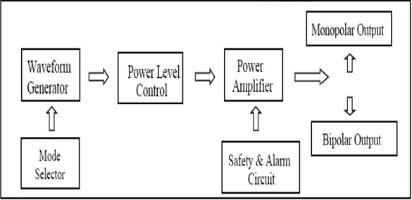
High-frequency power is required to generate heat from the waveform generator. Different waveforms like cutting, coagulation and blending are also produced from the generator. The mode selector is used for selecting which type of mode should be used while performing the surgery. Power level control determines the power applied to the electrodes during surgery.  Alarm circuit is used for creating safety alarms whether the electrodes are attached properly in the patient circuit. 

Circuit Components


The front panel consists of:- 
Power ON/OFF switch – Turning On/ Off of the electrosurgical unit. 

Mode selector – For selecting the modes for performing surgery (CUT mode, COAG mode, BLEND mode ). Cut mode indicates a yellow colour, whereas Coagulation mode indicates a blue colour. 

Fault indicator switch – This indicates poor contact of patient return electrode with the patient by producing alarm. Three-pin switch for monopolar use only hand switch. Three-pin switch for bipolar can be activated with or without footswitch. There is also a two-pin connector for the patient return electrode.
Rear panel Consists of:-
Footswitch control – This is for connecting the footswitch. 

The volume control knob - is used for controlling the audio volume during cutting, coagulation or blending. Power cord connection.

Safety precautions
  • Electrosurgery must be performed by specially trained technicians in electrosurgical technologies.
  • Electrosurgery should not be performed in oxygen-rich environments because it may easily catch fire and cause serious burns. 
  • Before operating, insulation of the electrode must be checked carefully to avoid thermal injuries. 
  • Do not activate the equipment in an open circuit.

Do’s
Electrodes should be placed in clean, dry areas
Use only insulated electrodes
Electrodes must be placed in well-cleaned holster, when not in use
Electrode tips must be cleaned properly.
Use only low voltage waveforms while operating
Bipolar electrosurgery can be performed when necessary

Don’ts
  • One should not use the electrode in the presence of flame causing agents like (alcohol, tincture etc.) to avoid burns and fire.
  • Electrodes should not be wrapped around metal instruments because they cause burns.
  • Rubber catheters should not be used in electrode tips.
  • One should not operate the electrosurgical unit by standing on a wet surface to prevent them from shocks.
  • Surgeons or technicians must not perform the operation with wet hands/wet gloves to prevent electric shock.
  • Must wear surgical masks during surgery to avoid smoking difficulties which may cause breathing problems.
  • Avoid using the electrode near electric devices.
  • Do not use the equipment in patients prior to testing. 

Problems in ESU
If surgery is performed in oxygen-rich environments, it may cause a surgical fire.
Patient burns may also be caused by detachment of the electrode pad.
Incorrect placement of the electrode may also cause burns.
Electrosurgical injury occurs when the electrode comes in contact with non-target tissue.
Since the device uses high current, it may cause electric shock, the transmission of infection etc.
Direct skin contact will cause burns.

Preventive Maintenance
  • Regular testing of the equipment should be done to prevent any harm and to enhance patient and staff safety. 
  • The person handling the equipment must be well trained by the manufacturer.
  • He/She must follow the instructions given in the manual. 
  • The technician must carefully inspect the insulation. 
  • Lower power setting must be used when starting the equipment for use.

Risks of Electrosurgery
  • Most of the risks are caused by thermal injuries. This is mainly due to unintended contact of the active electrode with any part of the body tissues. 
  • Another kind of thermal injury is caused by the contact of the active electrode with metal instruments.
  • Injuries caused by smoke also causes breathing problems in both patients and surgeons.
  • Major risks are associated with respiratory illness and irritation in the eyes, skin etc.
  • Therefore, proper measures must be taken before handling the equipment during surgery.

Electrical safety
  • The proper power cord must be used. It must be properly grounded.
  • Use only proper fuses which have correct voltage and current rating.

Patient Safety
Proper contact must be maintained between the patient and other metal parts to avoid burns.
Patient electrode must be connected in order to avoid skin injuries.
Ensure the connections made do not affect the blood supply in the patient.

Clinical Applications
  • Electrosurgery is often used in most of the operations involving tissue surgeries
  • ENT (To control nose bleeding)
  • Gynaecology
  • Ophthalmic surgery
  • Dermatology (To remove abnormal skin growth, moles etc.)
  • To remove lesions, hair follicles.
  • Also used in surgeries associated with organs like liver, spleen, kidneys and heart surgery.

Environments of use
Electrosurgery is used in operating rooms in the hospital while performing surgeries.

Troubleshooting
  • Check Electrosurgical unit for any physical damage
  • Ensure all cables, electrodes, power cord is connected properly
  • Check the power cord properly or immediately replace it.
  • Pin connections and switches must be properly checked. If any damage is found, it must be replaced immediately.

👉 Watch ESU Video on Our YouTube Channel Below:- 


Article Prepared By:-
👉 Sumitha Ramadass


References
  • https://www.ncbi.nlm.nih.gov/pmc/articles/PMC3407433/#:~:text=Principles%20of%20Electrosurgery,Often%20%E2%80%9Celectrocautery%E2%80%9D%20is&text=Electrocautery%20refers%20to%20direct%20current,alternating%20current%20(Figure%201).&text=Electrical%20current%20flows%20when%20electrons,produced%20when%20electrons%20encounter%20resistance.
  • http://www.boviemedical.com/2016/10/03/bipolar-electrosurgery-vs-monopolar-electrosurgery/
  • https://www.asit.org/assets/documents/Prinicpals_in_electrosurgery.pdf
  • https://www.glowm.com/section_view/heading/electrosurgery-principles-biologic-effects-and-results-in-female-reproductivesurgery/item/21#:~:text=There%20are%20three%20basic%20types,%2C%20nonmodulated%20sinusoidal%20waveform%20(Fig.
  • https://veteriankey.com/surgical-modalities-laser-radiofrequency-ultrasonic-and-electrosurgery/
  • https://www.semanticscholar.org/paper/Electrosurgery%3A-principles-and-practice-to-reduce-Brill/ba1a6e6b411cba18601762375b62562cdf166677/figure/5
  • https://www.uptodate.com/contents/overview-of-electrosurgery
  • https://www.slideshare.net/prasadvagal/electrosurgical-unit
  • https://www.ncbi.nlm.nih.gov/pmc/articles/PMC4579996/#:~:text=An%20electrosurgical%20unit%20(ESU)%20consists,do%20the%20corresponding%20tissue%20effects.
  • https://avantehs.com/p/11947
  • https://www.cehjournal.org/article/electrosurgical-units-how-they-work-and-how-to-use-them-safely/
  • https://www.gimaitaly.com/prodotti.asp?sku=30512&dept_selected=44&dept_id=446
  • http://www.micromed.com/en-US/products/electrodes/
  • https://www.sutter-med.de/en/products/monopolar-instruments/monopolar-electrodes_aid_50.html
  • https://www.who.int/medical_devices/innovation/electrosurgical_unit.pdf
  • https://www.flukebiomedical.com/blog/what-are-electrosurgical-units-why-should-we-test-them%3F
  • https://webstor.srmist.edu.in/web_assets/srm_mainsite/files/downloads/esu.pdf
  • https://www.magmedical-equip.com/product/valleylab-force-fx-c-electrosurgical-unit/
  • https://docplayer.net/21481453-Instructions-electrosurgical-unit-psd-30.html

PageNavi Results Number

Contact Us via Email to Know More About Our Supports...:- sam.gastondiaz@gmail.com