TYPES OF ENDOSCOPES AND THEIR USAGE:
- ARTHROSCOPE: The procedure of arthroscope is known as arthroscopy. The arthroscope is specially designed for the area of joints, it put in through cuts in the skin.
 |
| IMAGE OF ARTHROSCOPE |
- BRONCHOSCOPE: The procedure of bronchoscope is known as bronchoscopy or flexible bronchoscope. It is specially designed for the area of trachea (windpipe) and bronchi (tubes going to the lungs), it put in through mouth or nose.
 |
| IMAGE OF BRONCHOSCOPE |
- COLONOSCOPE: The procedure of colonoscope is known as colonoscopy or lower endoscopy. It is specially designed for the area of colon and large intestine, vagina and cervix, it put in through anus.
 |
| IMAGE OF COLONOSCOPE |
- CYSTOSCOPE: The procedure of cystoscope is known as cystoscopy or cystourethroscopy. It is specially designed for the area of the bladder, it put in through urethra.
 |
| IMAGE OF CYSTOSCOPE |
- ENTEROSCOPE: The procedure of enteroscope is known as enteroscopy. It is specially designed for the area of the small intestine, it put in through mouth or anus.
 |
| IMAGE OF ENTEROSCOPE |
- ESOPHAGOGASTRODUODENOSCOPE: The procedure of esophagogastroduodenoscope is known as esophagogastroduodenoscopy(EGD), upper endoscopy, panendoscopy or gastroscopy. It is specially designed for the area of the oesophagus (swallowing tube), stomach and duodenum (first part of the small intestine). It put in through the mouth.
 |
| IMAGE OF GASTROSCOPE |
- HYSTEROSCOPE: The procedure of hysteroscopy is known as hysteroscopy. It is specially designed for the area of the uterus, it put in through the vagina.
 |
| IMAGE OF HYSTEROSCOPE |
- LAPAROSCOPE: The procedure of laparoscope is known as laparoscopy or peritoneal endoscopy. It is specially designed for the area of the abdomen and pelvis, it put in through small incision in the abdomen.
 |
| IMAGE OF LAPAROSCOPE |
- LARYNGOSCOPE: The process of the laryngoscope is known as laryngoscopy. It is specially designed for the area of the larynx (voice box), it put into the mouth or nose.
 |
| IMAGE OF LARYNGOSCOPE |
- MEDIASTINOSCOPE: The process of mediastinoscope is known as mediastinoscopy. It is specially designed for the area of the mediastinum(space between the lungs), it put through the incision above the breastbone(sternum).
 |
| IMAGE OF MEIASTINOSCOPE |
- SIGMOIDOSCOPE: The process of sigmoidoscope is known as sigmoidoscopy or flexible sigmoidoscopy or proctosigmoidoscopy. It is specially designed for the area of the rectum and sigmoid colon (lower part of the large intestine), is put through the anus.
 |
| IMAGE OF SIGMOIDOSCOPE |
- THORACOSCOPE: The process of thoracoscope is known as thoracoscopy or pleuroscopy. It is specially designed for the area of the space between the lungs and chest wall, it put through the incision into the chest cavity.
 |
| IMAGE OF THORACOSCOPE |
IS THERE ANY STERILIZATION PROCEDURE IS THERE?
Endoscopes are routinely exposed to mucus and other gastrointestinal secretions, blood, saliva, faeces, bile and sometimes pus.
- Autoclaving is an economic and excellent method of sterilizing the instruments that do not heat sensitive. If the instruments are autoclaved using hot sterilization methods the heat-sensitive instruments (flexible ureteroscope, chip on tip endoscopes) get damaged.
- Manual cleaning includes brushing with single-use wire brushes and exposure of all external and accessible internal components to a low-foaming enzymatic detergent.
- Chemical cleaning includes glutaraldehyde, hydrogen peroxide, peracetic with hydrogen peroxide are used.
- A new method of sterilization by moist heat has been used in the laboratory. It involves immersing the contaminated instrument is a water bath at 85°c for one hour. This becomes possible for heat-resistant. It reported that it killed most pathogenic organisms including mycobacterium tuberculosis, E.coli, Proteus Vulgaris and pseudomonas pyocyanin.
IMAGES TAKEN DURING GASTROSCOPY:
ADVANCES IN ENDOSCOPES:
- Advances in digital imaging provide the viewer with a more engaging, immersive experience. In digital imaging, Endoscopists are becoming increasingly able to detect subtle, minute mucosal changes that were previously indistinguishable from normal tissue.
- Advances in television, the endoscope has switched from “standard definition” to digital, high-definition white light imaging. It results in image enhancement.
- The endoscopy has begun to play an ever-increasing role in the detection and treatment of gastrointestinal cancers, oesophagal cancers and other GI conditions.
- Ability to treat not only the mucosal disease but also the submucosal disease.
- Narrowband imaging (NBI) introduced in endoscopy for early detection of lesions, this shows microvascular pattern which allows better characterization of lesions.
- The use of HD endoscopes and monitors allows substantial image improvements by producing fewer artefacts on rapid movement and when combined with corresponding processors may reach an image quality of over 2 million pixels.
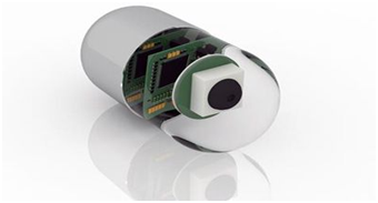
CAPSULE ENDOSCOPY:
 |
| IMAGE OF CAPSULE ENDOSCOPE |
 |
| IMAGE OF CAPSULE ENDOSCOPY INSIDE THE STOMACH |
Capsule endoscopy is a procedure that uses a tiny wireless camera to take pictures of your digestive tract.
- A capsule endoscopy camera sits inside a vitamin-size capsule you swallow.
- As the capsule travels through your digestive tract, the camera takes thousands of pictures that are transmitted to a recorder you wear a belt around your waist.
- It also helps to view the area that isn’t easily reached
- It is mostly done when the cause of gastrointestinal bleeding, cancer diagnosis, polyps screening etc...
RISK AND SIDE EFFECTS OF ENDOSCOPY:
- Reaction to sedation prior to your upper endoscopy some sedation has to be given after the procedure, sometimes it causes the adverse risk of a serious reaction.
- Sometimes tearing of the gastrointestinal tract may occur.
- Mostly endoscopies consist of an examination and biopsy and so the risk of infection is low. When the risk of infection increases when additional procedures are performed.
- If the biopsy has been done then the risk of bleeding complication may occur. In rare cases, such bleeding may require a blood transfusion.
SIGNS AND SYMPTOMS AFTER ENDOSCOPY:
- Vomiting
- Fever
- Chest pain
- Difficulty in swallowing
- Shortness of breath
- Persistent abdominal pain
- Bloating and gas
- Cramping
- Sore throat
ABNORMALITIES DETECTED DURING ENDOSCOPY:
- Mucosal abnormalities are detected such as erythema, ulcer, erosion and polyp.
- Narrowed areas and strictures of the oesophagus, stomach or duodenum are detected and treated.
- Objects stuck in the oesophagus or stomach can be detected and removed.
- Bleeding due to ulcers can be detected.
- Precancerous abnormalities such as Barrett’s oesophagus is detected.
- Inflammation or swelling can be detected.
- Blockages are detected.
- Gastroesophageal reflux disease ( it is a digestive disease in which stomach acid or bile irritates food pipe) is detected, for this biopsies are needed to diagnose.
HOW TO MAINTAIN ENDOSCOPE:
Maintenance is very important for medical equipment.
- Pre-cleaning is important to clean priorly for the disinfection after the procedure done immediately wipe the endoscope using an approved enzymatic solution. Wash suction, air and water channels according to the manufacturer’s instruction.
- Always test the endoscope for leaks, immerse in water and test.
- Thoroughly rinse the exterior of the endoscope using clean water.
- Clean all channel with proper disinfectant.
- Use alcohol-water mixture (70 to 80 percent ) to flush all, then wipe and dry.
- Store the endoscope in a well-ventilated container.
TROUBLES IN ENDOSCOPE:
- The left-hand holds the control section with the right hand controlling the insertion tip, be aware of kinking of the upper end of the umbilical cord. If it changes during bending it could cause damage in the internal channel.
- The most proximal portion of the endoscope is the eyepiece. The generic replacement eyepiece commonly repairs by unauthorized repair firms in which they use low-quality materials so during the examination it may cause an undesirable view.
- Sometimes the shaft (it contains all illumination components) in the endoscopes may get damaged.
HOW TO TROUBLESHOOT THE PROBLEMS:
- Correct positioning for each procedure results in the safest operation/endoscopy.
- The high-quality standard authorized eyepiece is fitted according to the manufacturer’s specification.
- Always use the endoscopes with proper guidance.
👉Please Watch Our Endoscope (Part 2) Video From Our YouTube Channel Below:-
REFERENCE:
1.
https://www.sciencedirect.com/topics/nursing-and-health-professions/gastroscope
2.
https://www.ncbi.nlm.nih.gov/books/NBK310264/
3.
https://www.nhs.uk/conditions/gastroscopy/what-happens/
4.
https://link.springer.com/chapter/10.1007/978-4-431-78889-8_1
5.
https://www.bupa.co.uk/health-information/digestive-gut-health/gastroscopy
6.
https://www.olympus-global.com/technology/museum/endo/?page=technology_museum
7.
https://www.docdoc.com.sg/info/procedure/gastroscopy/


No comments:
Post a Comment